A Muscle Watch: A Personalized Device for Hamstring Injury Prevention Using Surface Electromyography (sEMG) Sensors and Machine Learning
Grade 9
Presentation
Problem
Motivation
- “Hamstring muscle is the most common muscle strain injury in high-speed running sports [1]. Injuries acquired during maximal velocity running make up 47% of all hamstring injuries, accounting for 24% of all injuries in soccer, 17% in track and field [2]. Upon Return to Play (RTP), the injury reoccurrence rate ranges from 18% to 33% [2, 3]. Athletes are 4.8 times more likely to sustain a hamstring injury if they had previously suffered a hamstring injury within the last season [3].
- Not only do hamstring injuries result in lengthy amounts of time off from sport [3], reduced physical activity, and economic costs [4], but they also affect the injured athletes psychologically 100% of the time as reported by two-thirds of Canada-based sports chiropractors in a 2023 study [5]. Adolescence, being in a stage of transformative physical, social, and psychological development [6], are more vulnerable to experiencing consequences of injury such as depression, anxiety, a loss of confidence and a growing sense of doubt [7]. Lifelong consequences may impact one’s quality of life, activity level in adulthood, and the possible development of mental health disorders [6].
- Canadian youth are at risk of hamstring injuries given:
- Their high participation rates (86.89% in 2019) in basketball, badminton, soccer, volleyball, and running [8], sports that involve frequent changes of direction, pivoting turns, landings [9] and sprinting [1]; and
- A lack of awareness of the risk or prevention strategies, particularly players who do not have access to professional coaches or trainers in urban team sport settings [4].
- Current hamstring injury prevention measures such as eccentric strength training [1, 3] and the SHRed Neuromuscular Training warm-up programs developed by the University of Calgary Sport Injury Prevention Research Centre (SIPRC) have shown to be effective at reducing injury rates when implemented in youth team sports [9]. However, these strategies only prevent injuries with adequate program adherence (i.e. use as intended by players and coaches) [10, 11].
- Even when program adherence at the team level is satisfactory, monitoring an individual’s exercise fidelity remains challenging [12, 11]. Exercise fidelity is a measure of how well an athlete performs the exercises “with the correct technique and intensity and completing the prescribed sets and repetitions.” [9]
Question
How might we non-invasively monitor hamstring muscle activation in teenagers during physical activity to prevent hamstring injury and minimize downtime in play?
Project Objectives
I am proposing the creation of a personalized, wearable leg sleeve device equipped with surface electromyography (sEMG) sensors and machine learning (ML) to provide real-time feedback (based on ML predictions) on the state of hamstring muscle activation. This innovative technology (which I will refer to as the MuscleWatch) is intended to be used as a real-time feedback tool for improving youth athletes’ exercise fidelity when used in conjunction with the current, clinically proven hamstring injury prevention strategies and/or hamstring injury rehabilitation programs.
There are two groups of prospective users who will benefit from the use of this MuscleWatch:
- Youth athletes who are active in play: the device serves as a monitoring tool alongside current hamstring injury prevention strategies in minimizing downtime in play.
- Injured youth athletes who are undergoing rehabilitation: the device serves as a feedback tool for adjusting sport-specific loading of the hamstring muscles to speed up progression towards Return to Play and/or prevent re-injury.
This project has three phases:
- Design a personalized, wearable technology sEMG sensing device using consumer-level hardware components.
- Develop all the MuscleWatch components:
- the physical leg sleeve,
- the sEMG signal acquisition module which includes the digital pre-processing logic (e.g. Arduino C++ code), and
- the microprocessor receiving module which includes the machine learning models to predict exercise fidelity adherence and the calculation of the predicted Rate of Perceived Exertion (RPE).
- Validate the feasibility and effectiveness of the MuscleWatch in improving one’s exercise fidelity, when used with current Neuromuscular Training (NMT) warm-up strategies, and thereby contributing to hamstring injury prevention in a simulated NMT warm-up environment.
- For the purpose of this science fair project, the scope of the validation is limited to the first group of prospective users, namely young athletes who are active in play.
Background Research
A. Hamstring muscles
- MuscleWatch will use two sEMG sensors to collect data from the medial region of the user's two hamstring muscles, the biceps femoris (BF) and the semitendinosus (ST) as illustrated in Figure 1. The biceps femoris and the semitendinosus were chosen as these muscles are the most superficial to the skin and thus have a larger surface area to place each sensor on [14].
- Additionally, the hamstring muscles are especially susceptible to injury, particularly in the BF due to heavy loading condition, and the greatest mechanical stress [1] during sprinting where the hamstrings must contract and release rapidly [9].
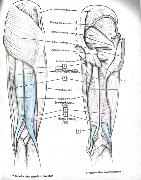
Figure 1. Coloured anatomy of the hamstring muscles (Sourced from: Netter’s anatomy coloring book [13]).
B. Wearable technology
- Wearable technology is described as non-invasive devices worn on the body which include electronic components such as sensors, processing units, and power sources [15].
- Wearable technology is popularized in sports as its ability to improve training and/or athletic performance and enhance safety [15].
C. Personalized device
- Personalized devices allow for more customizability particularly in:
- The fitting of the leg sleeve to ensure a correct placement of the sEMG sensors on the BF and ST muscles and a comfortable user experience.
- Machine learning that can identify trends and patterns in an individual’s exercise fidelity, offering personalized predictions and recommendations for improvement on hamstring muscle activation.
- Tailoring wearable technology devices to an individual user’s specific risk factors -- such as age, gender, muscle conditions, and previous injury -- can more effectively reduce the risk of hamstring injuries [2].
D. sEMG sensors (electrodes and hardware preprocessing)
- sEMG sensing is a proven, non-invasive physiological and quantitative assessment of muscle action potentials during muscle contractions [16].
- sEMG sensors offer the benefit of being portable while providing simple-to-understand and real-time measurements of muscle activation. This meets my design objectives of wearability and functionality (noted in the Method section below).
- Dry electrodes were chosen due to the following factors:
- No gels or conductive adhesives are required for placement on the skin.
- Metal electrodes are extremely durable and long lasting [17].
- I chose to use a commercially available sensor (DFRobot/OYMotion SEN0240) for these reasons:
- To stay within the project budget (see the Materials & Cost section below).
- It offers a plug-and-play experience that is compatible with Arduino analog pins [17].
- The sEMG signal is pre-processed within the sensor and its amplifier board where the raw sEMG signals (in microvolts) are extracted into time-domain, Analog-to-Digital-Converter (ADC) values [18]. These values can then be easily fed into the microprocessor receiving module for subsequent machine learning prediction.
E. sEMG signal extraction
- The 10-bit ADC values are transformed through various processes during signal extraction. These processes, as illustrated in Figure 5, include:
- Notch & Highpass filtering:
- Incoming ADC values are notch filtered to remove 60Hz powerline noise and highpass filtered to remove all noise below 20Hz [19].
- Thresholding:
- Filtered ADC values are rounded to zero when below a certain threshold to clearly show the difference between muscle activation and no muscle activation.
- Root Mean Square (RMS) [16]:
- Every 4 values are grouped into a window, and RMS is preformed resulting in an overall smoother transition between periods of muscle activity and no muscle activity.
- Down Sampling [20]:
- Post-RMS values are down sampled meaning 1 in every 8 values is taken to avoid passing on too much information to the Microprocessor receiving module.
- Notch & Highpass filtering:
F. Microcontroller configuration
- The Arduino Uno R4 was chosen as the microcontroller of the sEMG signal acquisition module for two reasons:
- It is compatible with the sEMG sensors made by DFRobot & OYMotion.
- It was the only Arduino board with both Bluetooth capabilities and 2 power pins for the 2 sEMG sensor connections.
- The Raspberry Pi was chosen for the microprocessor receiving module as it is a micro scale Linux computer, capable of running software designed for full-scale personal computers. This provided me with full access to the Jupyter lab code notebook and Tensorflow machine learning framework.
G. Machine learning (ML)
- Supervised learning, which is a style of machine learning, is used in this project where the models are fed with lots of examples to learn the relation between sEMG ADC values and the label of proper exercise fidelity [21].
- Binary classification models are designed to classify the input examples and generate a Boolean prediction – such as either “proper” or “improper” [21].
- The design and development choices used in my machine learning models are documented in the Method: Machine Learning Models Design and Development section below.
Method
Part I: MuscleWatch Design Objectives
1. Wearability:
- a lightweight, adjustable sleeve that can comfortably fit around a user’s leg
- sEMG sensors correctly placed according to SENIAM recommendations
2. Functionality:
- to provide a simple and quantitative assessment of the current state of the hamstring muscle activation
- seamless sEMG signal acquisition, signal processing/extraction, and wireless transmission of data to be processed and displayed to the users
- programming logic to determine when Neuromuscular Training (NMT) exercise fidelity is achieved according to the training dataset
- reliable power management
3. Materials & Cost:
- use consumer-level hardware components
- access to common household tools, a PC and a 3D printer
- a budget of $500
MuscleWatch Design Overview
- Adopting the basic wearable technology architecture, the MuscleWatch consists of 3 layers, as illustrated in Figure 2: sensor, processing, and network communication [15].
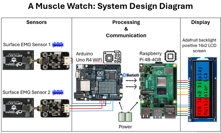
Figure 2. Hardware design diagram.
MuscleWatch Components
- Sensors: 2 sEMG sensors to be placed on the Semitendinosus (ST) and the Biceps Femoris (BF). The sEMG electrodes detect the electrical signals originating from these two hamstring muscles (ST and BF) that are measured in microvolts.
- Microcontroller: Arduino Uno R4 Wifi - takes sEMG ADC values and applies a 60Hz notch filter before relaying the data to the Raspberry Pi.
- Processing unit: Raspberry Pi 4B – run values received from the Arduino through the machine learning model via Jupyter lab.
- Communication unit: Bluetooth Low Energy (BLE).
- Display unit: RGB backlight positive LCD 16x2 display.
- Power units: 2 power banks (one for Arduino and one for Raspberry Pi).
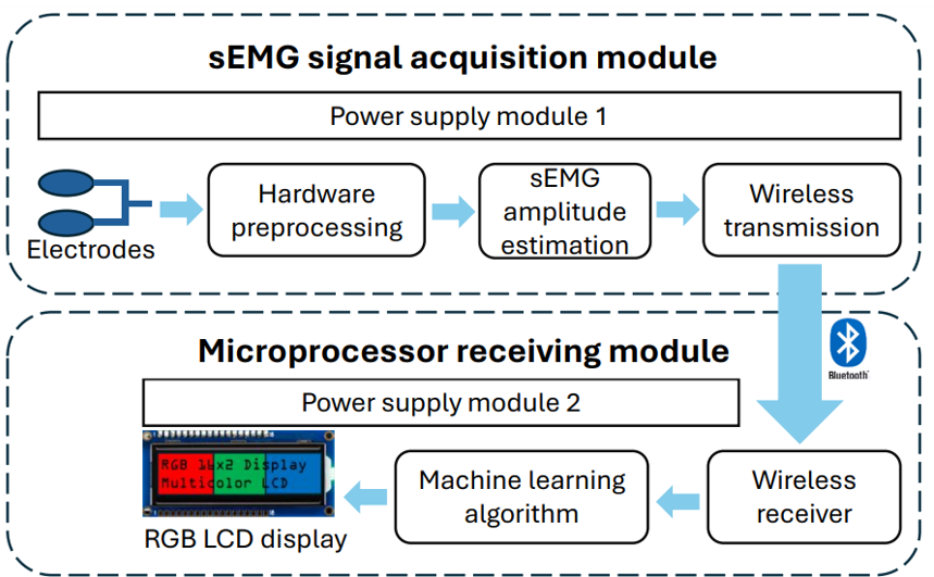
Figure 3. Block diagram of the MuscleWatch [16].
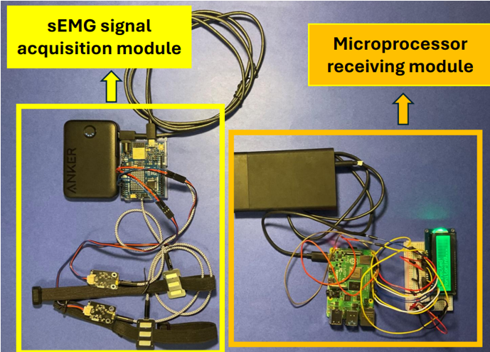
Figure 4. Physical diagram of the MuscleWatch [16].
Part II: MuscleWatch Development Requirements
Material & Cost
- All the materials that had to be purchased specifically for this project are listed in Table 1 below (left); Existing household tools, computing hardware and software that I have access to are listed in the right column in Table 1.
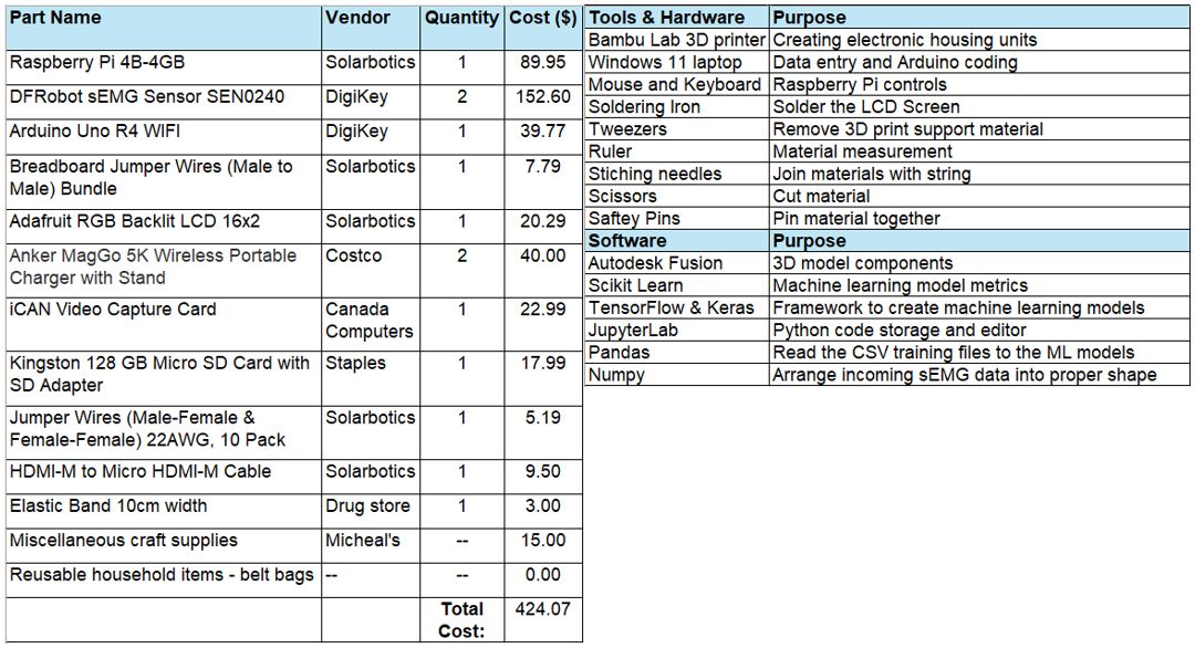
Table 1. Hardware, tools and software used to develop the MuscleWatch.
Hardware & Software Logic
- Figure 5 shows the process flow of the sEMG data throughout the signal acquisition module [18, 22].
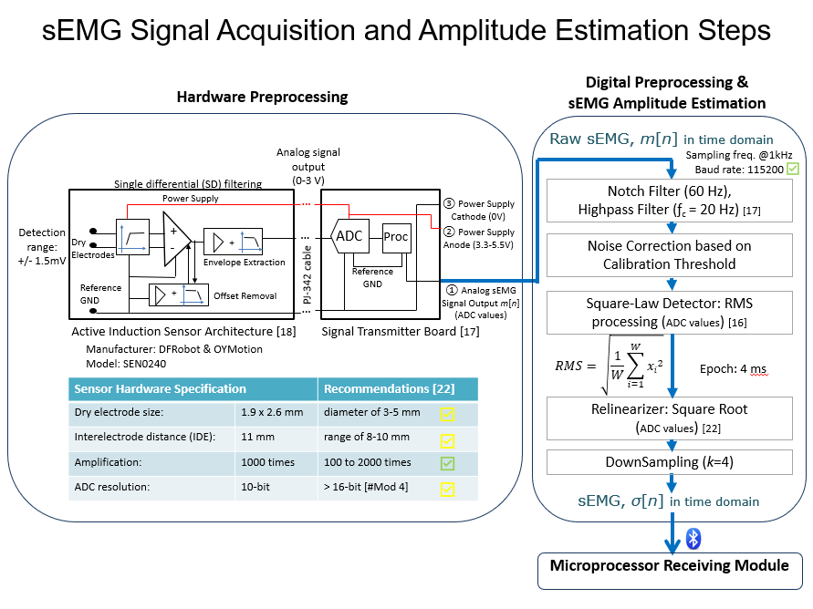
Figure 5. Hardware and Software preprocessing flowchart.
Microprocessor Receiving Module
- Refer to the section below titled “Machine Learning Models Design and Development” for the programming logic coded in this module.
3D Models for the Casing of MuscleWatch Hardware
- Four of the five 3D custom models, illustrated in Figure 6a, were created in Autodesk’s Fusion CAD software to house and protect the electronic components of the MuscleWatch.
- The other 3D model (located in the top middle of Figure 6a) was also designed in Fusion. It was engineered specifically to hold the fabric elastic bands with tension against the leg sleeve fabric so that the sEMG sensors could be moved along the four elastics anywhere within an xy-coordinate plane across the leg sleeve, like a train-track system, as shown in Figure 6b.
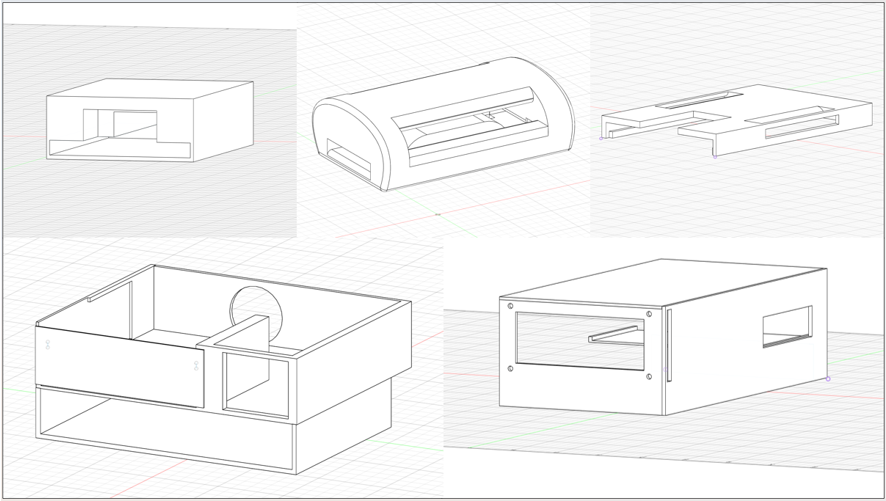
Figure 6a. Five 3D CAD models created in Autodesk Fusion used to 3D-print the electronic housing components.
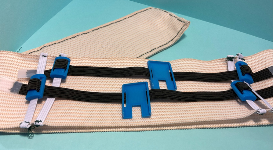
Figure 6b. Adjustable sEMG sensors on a train-track system in the MuscleWatch leg sleeve.
Machine Learning (ML) Design Objectives
- The speed of ML prediction should not interfere with the flow of the Nueromuscular Training (NMT) exercise repetitions (ideally it should be completed in seconds).
- The accuracy of each model should be greater than 50% to minimize misinterpretation of the actual muscle activation state during a repetition of an NMT exercise that is completed with proper exercise fidelity.
Machine Learning Models Design and Development
1. Problem
-
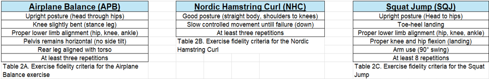
A binary classification is trained via supervised learning to predict whether exercise fidelity is met for each of the three NMT exercises:
- APB is the model for Airplane Balance;
- NHC is the model for Nordic Hamstring Curl;
- SQJ is the model for Squat Jump.
- Input variables are the 10-bit ADC values that are acquired from the sEMG signal acquisition module (sEMG sensors & Arduino).
- Classified output should either be put as “Proper” or “Improper” to indicate whether exercise fidelity has been met according to the assessment criteria [9] listed in Table 2 below.
2. Data collection and preparation
- Data is collected when I repeatedly perform each exercise 100+ times.
- Each ML example (think of it as a conventional row of data in a table) represents the sEMG ADC values acquired over one repetition of a NMT exercise in the range of 5 to 10 seconds depending on the NMT exercise.
- While training each of the ML models, I performed the first half of the examples in each dataset with proper exercise fidelity and then performed the second half with improper exercise fidelity (such as doing a very weak repetition or performing the motion incorrectly with little/no hamstring activation).
- By preparing the datasets in two separate portions ("proper" first and then followed by "improper" examples), I also got a better understanding of what the sEMG ADC data for an example with proper exercise fidelity looks like. This helps me identify and remove questionable examples that may affect the model training accuracy.
- Data is labelled based on my interpretation of whether proper exercise fidelity is met for each ML example.
- ML examples in the ML datasets are assigned a label of “Yes” (exercise fidelity criteria is met) or “No” (exercise fidelity criteria is not met).
- Data is preprocessed by shuffling examples in each dataset before ML training begins to prevent overfitting.
- If the examples are not shuffled given how the datasets were created in two portions as explained in the above, then it is likely that the Machcine Learning algorithm will be inaccurately training the given ML models. This is becuase the modesl will be constantly fed with "proper" examples one after another repeatedly until it reaches the second portion of the dataset where it will similarly be fed with only "improper" examples.
- Errors are removed and missing values are filled in for each applicable example in a dataset.
- For instance, ML examples that are visibly contaminated with noise (such as those that show identical values in the input variables) are removed from the dataset.
- To determine the number of hidden nodes needed, I divided the number of input variables in half to prevent overfitting across all three models: APB, NHC, SQJ.
3. Model selection
- My fully connected neural network has one hidden layer and one output layer.
- A sequential Keras neural network model is used due to my choice of using one input tensor and one output tensor.
- Keras default settings are used to initialize the weights.
4. Model training, evaluation and optimization
- Optimizer Stochastic Gradient Descent (SGD) is used to continually update the weight during the training of each model: APB, NHC, SQJ.
- Sigmoid activation is used for both layers across each model (APB, NHC, SQJ) as it puts the value to a range from 0 to 1 and pushes the output clearly to one class or the other. "ReLU" (rectified linear unit) activation was attempted but did not yield more accurate results.
- Scikit-learn metrics are used to generate each of the model’s loss and accuracy score on the validation and testing datasets throughout the training of all three models: APB, NHC, SQJ (refer to the confusion matrices illustrated in Table 3 below).
- Refer to the attachment “MLModels-TuningLog.pdf” to see the loss and accuracy through each iteration during model optimization.
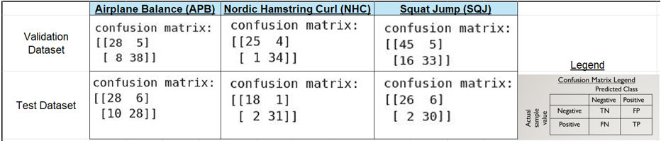
Table 3. Confusion Matrix of each machine learning model tested on a) the validation dataset (top row) and b) the test dataset (bottom row).
- Hyperparameters such as epochs, learning rate, batch size, and classification threshold are determined manually through trial and error due to non-linear hyperparameter interactions. The final hyperparameter values used for each model are listed in Table 4 below.
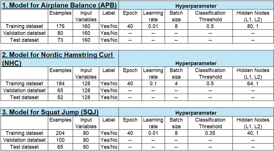
Table 4. Final ML Model Design Details and Hyperparameters for each of the three NMT warm-up exercises.
Prediction on the Rate of Perceived Exertion (RPE)
- The Rate of Perceived Exertion (RPE) was developed by Gunner Borg in the 1960s. RPE is used to measure how hard one’s body works during physical activity [23].
- The modified Borg CR10 RPE scale which runs from 0 to 10 will be used by the MuscleWatch (refer to Table 7 in Part III: Validation Study Objectives, in the Evaluation of the MuscleWatch section below).
- The MuscleWatch calculates the predicted RPE value using a preset maximum average value (that is specific to each participant), which is calibrated at the start of each simulation session.
- The preset maximum average values for the RPE calculations are calibrated before the simulation begins where I request the participant to perform a few repetitions of each of the NMT warm-up exercises while I monitor the ADC values coming from the sEMG sensors. During this time, I adjust the "maximum average value" based on my judgment to find the sweet spot in which the MuscleWatch displays "no activity" when the participant is not activating their hamstring muscles and displays an appropriately greater predicted RPE value when they begin to perform the exercises.
- The MuscleWatch’s predicted RPE is generated by calculating the percentage of the mean value (i.e. the average value calculated over a whole exercise repetition) over the individualized, preset maximum value as represented by this formula:
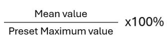
- This percentage-to-predicted RPE conversion is linear meaning the conversion happens as follows:
- 1-10% = a predicted RPE value of 1
- 11-20% = a predicted RPE value of 2
- 21-30% = a predicted RPE value of 3
- 31-40% = a predicted RPE value of 4
- 41-50% = a predicted RPE value of 5 and so on….
- For example:
- If the maximum mean value is set to 50 and the mean value across one entire Squat Jump repetition is 35, the calculated percentage will be 70% and thus gives a predicted RPE value of 7.
- To calibrate the MuscleWatch during simulation, I had to first ensure the participants were relaxed and standing (with minimal hamstring muscle contractions) and then wait for the MuscleWatch to produce a predicted RPE value:
- If the predicted RPE value was either a “0” or “1”, then I could procced with the next step of calibration.
- If the predicted RPE value was greater than 1, then I had to adjust the maximal mean value as needed to satisfy the previous condition.
- The second step of calibration was done while the participants were performing the NMT warm-up exercise. I had to check that the MuscleWatch was predicting higher RPE values than the prior RPE values when participants were stationary. Once a maximal mean value was found that can fulfill this delicate balance, the MuscleWatch is calibrated and ready for use.
Source Code
- All source code for the MuscleWatch can be found in my Github repository:
https://github.com/isaacC-2B/MuscleWatch-SourceCode
- Please read the README.md file to view the software copyright notices as the code development of the MuscleWatch involved some usage of generative AI, specifically ChatGPT and its adaption CS50.ai, which was developed by Harvard University to support students who are learning to code.
Part III: Validation Study Objectives
- To verify, post-simulation, the accuracy of the trained machine learning (ML) models by comparing the ML models (based on the sEMG data collected) with the visual assessment of exercise fidelity (based on video analysis using the exercise fidelity criteria [9], outlined in Figure 7 below).
- To determine if the provision of real-time biomechanical data during Neuromuscular Training (NMT) warm-up will improve an individual’s exercise fidelity (the volume and quality of hamstring muscle activation).
- To investigate the feasibility and effectiveness of the MuscleWatch in contributing to the current, clinically proven hamstring injury prevention strategies (such as the SPIRC-SHRed Injury Neuromuscular Training Warm-up Program) used in a simulated setting.
Workflow of the Validation Study
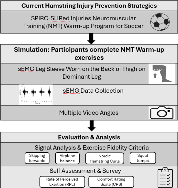
Figure 7. A workflow diagram of the key processes and methods used in the validation study in evaluating the MuscleWatch [9].
Participants
- 11 participants (mean ± SD age 13.5 ± 2.2) volunteered to participate.
- All participants (or their parents if under 18 years of age) provided written informed consent for participation in this study.
- Participants must have passed the 2022 PAR-Q+ assessment (The Physical Activity Readiness Questionnaire for Everyone) to participate in this study [24].
- Participants were recruited from my school’s athletics teams including but not limited to soccer, cross country, track and field, basketball, and badminton.
Simulation Design & Setup
- The simulation took place in the living room of my house where there is adequate space to perform the warm-up exercises.
- Participants were given an orientation on:
- how the MuscleWatch works;
- how to adjust the device for a better fit on their dominant leg; and
- what is the proper placement of the sEMG sensors.
- Participants were shown a video from the University of Calgary Sport Injury Prevention Research Centre (SIPRC) website [25], demonstrating each of the 4 Neuromuscular Training (NMT) warm-up exercises listed in Table 5 below.
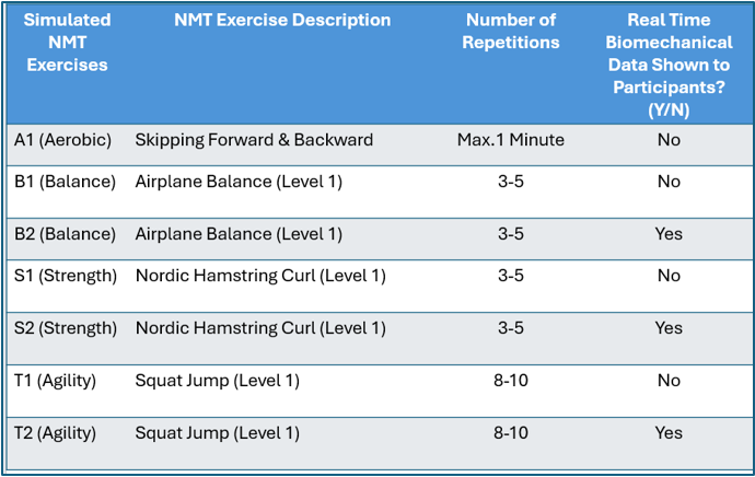
Table 5. Four NMT warm-up exercises performed by each participant.
- Participants performed the 4 common, low intensity, repetition based, NMT warm-up exercises for Soccer and Basketball as listed in Table 5 above. These 4 exercises (one from each of these categories: Aerobic, Balance, Strength, and Agility) were specifically selected for the concentrated focus on hamstring muscle activation.
- The Skipping Forward & Backward exercise, namely A1 (Aerobic) in Table 5, was performed at the start of the simulation where the participants’ hamstring muscles were being warmed up.
- Participants completed a Rate of Perceived Exertion (RPE) self-assessment.
- The modified Borg CR10 RPE scale which runs from 0 to 10 was used.
- Participants completed a Comfort Rating Scale (CRS) survey on their experience of comfort while wearing the MuscleWatch during the NMT warm-up simulation.
- The Comfort Rating Scale (CRS) was developed by Knight and Baber (2005) to assess wearer comfort across six dimensions for wearable technologies [26].
- The detailed step-by-step instructions for the simulation are documented in the Test Script (Table 6).
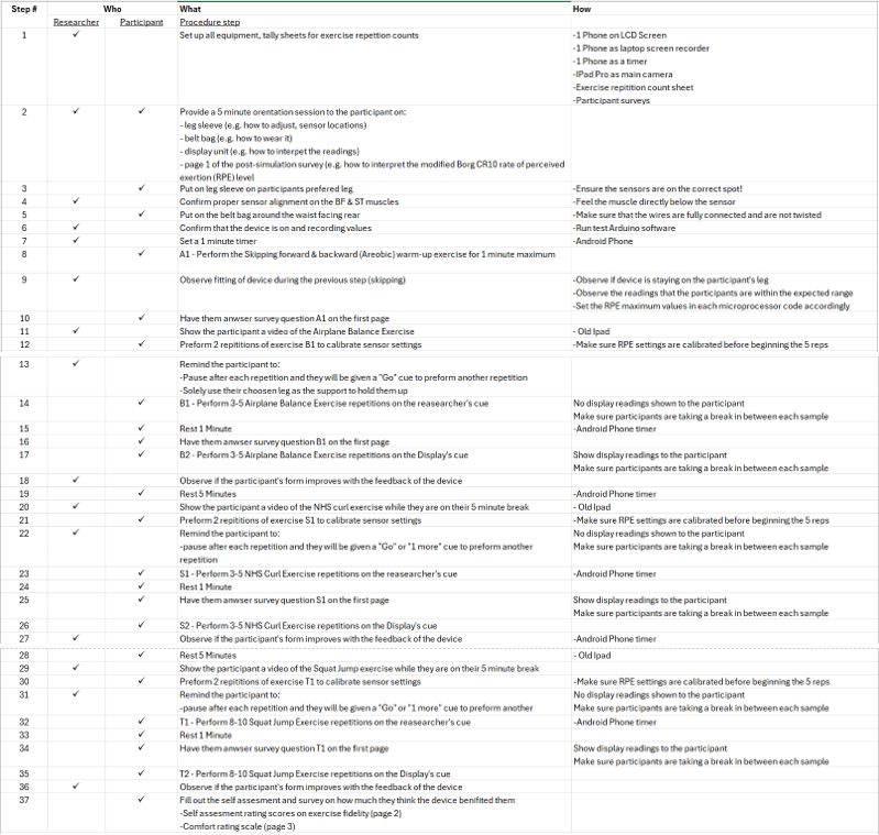
Table 6. Validation study test script used in the simulation for each participant.
Simulation Data Collection
- sEMG data of each participant’s hamstring muscle activation was measured during the exercises.
- A video log of the device’s output (the colour-coded biomechanical data readings shown on the MuscleWatch display unit) for every repetition of the exercises performed by each participant was recorded.
- A video of each exercise performed by some of the participants was recorded for subsequent analysis by me, as needed, post-simulation.
Evaluation of the MuscleWatch
The following tools were used at the conclusion of the simulation.
1. Participant self-assessment:
- Did the participant’s Rate of Perceived Exertion (RPE) align with the MuscleWatch’s prediction of hamstring activation? (Tables 7 & 8)
- Did the participants find having real-time hamstring muscle activation data useful? If yes, how were they positively influenced by using it in improving their warm-up exercise performance? (Table 9)
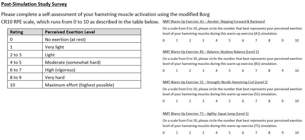
Table 7. The modified Borg CR10 RPE level. Table 8. Post-simulation RPE self-assessment.
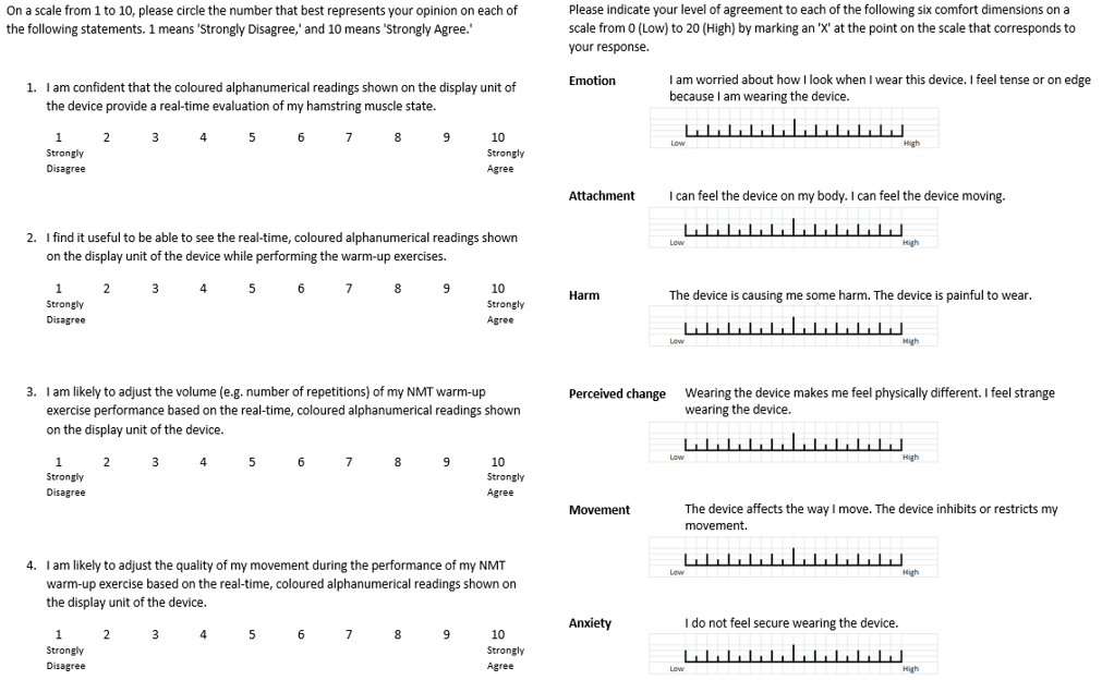
Table 9. Tool to assess the MuscleWatch. Table 10. Survey to assess Comfort Rating.
2. Survey
- Participants filled out a Comfort Rating Scale (CRS) survey (Table 10) to provide their judgement of comfort when wearing the proposed sEMG device.
3. Exercise fidelity criteria were assessed by the researcher
- Compared the MuscleWatch’s display readings for these two simulation scenarios to see if each participant’s exercise performance differed:
- without the MuscleWatch's readings shown to the participants (i.e. exercises B1, S1, and T1 listed in Table 5); and
- with the MuscleWatch's readings shown to the participants after each repetition of a NMT exercise (i.e. B2, S2, and T2 in Table 5).
- Compared the machine learning algorithm in detecting proper exercise performance based on hamstring muscle activation vs. human evaluation of the quality completion of the NMT warm-up exercise (based on exercise fidelity criteria) through direct observation and/or video analysis.
Analysis
Observations
- The total number of Neuromuscular Training (NMT) exercise repetitions, across all three exercises, preformed by the 11 participants, and analyzed was 569.
- Leg size and accessible hamstring muscle area played a large role in the length of time required to secure the MuscleWatch with the correct placement of the sensors.
- Achieving correct sEMG sensor placement required some additional contact pressure to hold the electrodes tight to the skin. An extra piece of 3D-printed backing had to be added to the back of the sensor plates on the leg sleeve for 4 out of the 11 participants.
- Calibration of both the Rate of Perceived Exertion (RPE) calculations and sEMG thresholds was needed for every participant and every NMT exercise as varying physiological and execution differences needed to be accounted for during both the signal acquisition and the RPE classification.
- The power units of the MuscleWatch remained stable and delivered a reliable supply of power to all the electronic components throughout each simulation.
- The MuscleWatch worked as intended where the display showed two different machine learning (ML) predictions for each repetition of an NMT exercise.
- Red (representing “improper”) or Green (representing “proper”) shows if exercise fidelity is upheld for the specific NMT exercise.
- The RPE value (on a scale of 0-10) shows a predicted exertion value.
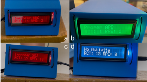
Figure 8. (a) An example of the MuscleWatch display showing its ML prediction of an improper NHC exercise repetition and a predicted RPE value of 2. (b) An example of the MuscleWatch display showing its ML prediction of a proper APB exercise repetition and a predicted RPE value of 1. (c) An example of the MuscleWatch display showing its ML prediction of an improper SQJ exercise repetition and a predicted RPE value of 10. (d) An example of the MuscleWatch display showing no ML prediction of exercise fidelity during an idle period of sEMG acquisition and hence a predicted RPE value of 0.
- The execution of repetitions (especially for the Airplane Balance Exercise) was noticeably inconsistent and varied from participant to participant, which resulted in a wide variety of muscle activation patterns and/or improper hamstring muscle activation.
- Many participants were ecstatic to see both the MuscleWatch predictions: “Proper” or “Improper” for exercise fidelity and the predicted RPE value.
Results & Analysis
- The results presented in this section are assessed against the validation objectives.
Objective #1: To verify the accuracy of the trained machine learning (ML) models.
- In all three ML models (shown in Figure 9), the validation accuracy on myself (the rightmost bar colored in dark blue) had the best result -- it was 10-20% more accurate than the predictions on the participants. This was anticipated given the training of the ML models was limited to acquiring sEMG data only from myself.
- Overall, the Nordic Hamstring Curl ML model showed the best accuracy and was the only exercise where participants’ exercise fidelity improved when the participants were shown the display of MuscleWatch readings.
- Both the NHC and SQJ model’s RPE prediction charts (Figure 10b) appeared to be more symmetrically mirrored, which suggests that exercises like the Airplane Balance, where muscles throughout the entire body are activated, could potentially introduce errors in the acquired sEMG hamstring data from the Biceps Femoris & Semitendinosus due to incorrect execution of the exercise and thereby adversely impacting the hamstring muscle activation.
- While the Airplane Balance and the Squat Jump both engage various muscle groups in the lower extremities, the Nordic Hamstring Curl has a primary focus on engaging the hamstrings, making it a repeatable and consistent motion across all repetitions.
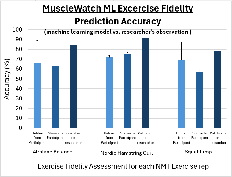
Figure 9. A comparison of the MuscleWatch machine learning prediction accuracy on exercise fidelity between the average value that was recorded for all participants during validation and the prediction accuracy results obtained during the machine learning model evaluation phase using my own validation dataset for each of the 3 NMT warm-up exercises.
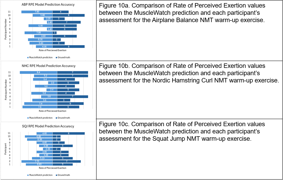
Figure 10. A comparison between the MuscleWatch RPE prediction accuracy and each participant’s self-assessment of their RPE during each of the 3 NMT warm-up exercises.
Objective #2: To determine if the provision of real-time biomechanical data during Neuromuscular Training (NMT) warm-up will improve an individual’s exercise fidelity (the volume and quality of hamstring muscle activation).
- Participants found it useful to learn of their real-time biomechanical data as shown in Figure 11.
- Most participants would prefer to adjust the quality of their NMT exercise repetitions (an average of 7 out of 10 rating) rather than the volume (an average of 5.3 out of 10) as shown in Figure 11.
- Most participants were confident that the MuscleWatch is accurately showing their current hamstring muscle activation state readings.
- The MuscleWatch prediction speed was satisfactory, coming up with a prediction every few seconds, which did not hinder the participant’s repetitions. As the video shows (timestamp at 1:42), a repetition of the Nordic Hamstring Curl followed by the MuscleWatch’s prediction completed in just under 9 seconds. (Note that each cycle of sEMG signal acquisition ranges from 5-10 seconds depending on the exercise).
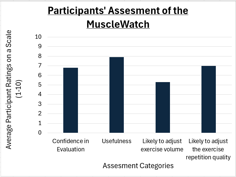
Figure 11. Participants’ confidence assessment of the MuscleWatch
Objective #3: To investigate the feasibility and effectiveness of the MuscleWatch in contributing to the current, clinically proven hamstring injury prevention strategies (such as the SPIRC-SHRed Injury Neuromuscular Training Warm-up Program) used in a simulated setting.
- As shown in Table 11 below, all ratings were within the range of Wearability Level 1 to Wearability Level 3 indicating that the MuscleWatch is wearable although improvements in attachment are recommended.
- The highest rating was attachment (12.9), however, its standard deviation (4.26) suggested that only a few participants felt that the MuscleWatch had high attachment which in turn raised the average value. I see this as an acceptable result given the sEMG sensors were not attached to the participants with any adhesives in following the CYSF low-risk project guidelines.
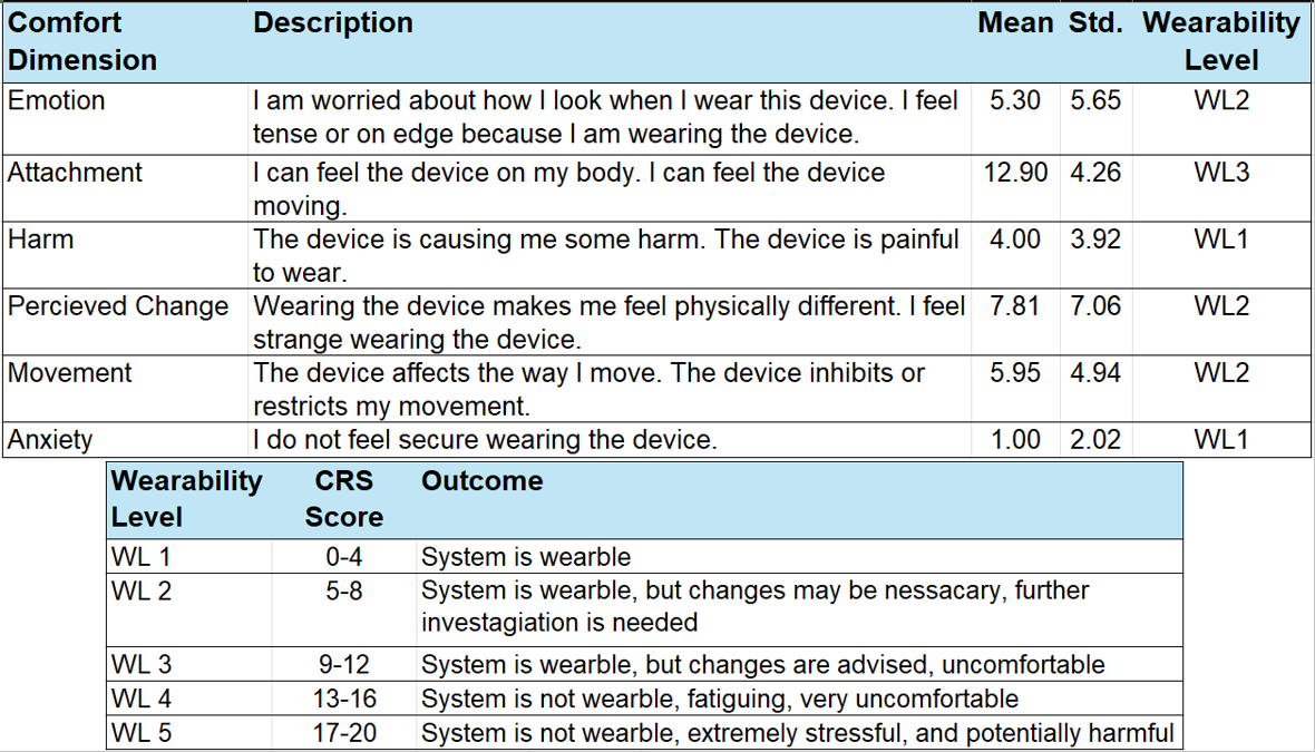
Table 11. Participants’ assessment of the Comfort Rating Scale of the MuscleWatch, along with the Wearability Level results (top); The meaning of each Wearability Level as proposed by Knight et al. [27] (bottom).
- As shown in Figure 12 (below) and Table 11 (above), the next highest field after attachment was perceived change (7.81), followed by movement (5.95). The results of these fields suggested that the MuscleWatch could be affecting the participant’s exercise fidelity, perceived RPE and possibly the accuracy of the MuscleWatch.
- The participants reported little to no discomfort, pain, or anxiety related to the use of the MuscleWatch with respective scores of a harm score: 4.00 and an anxiety score: 1.00.
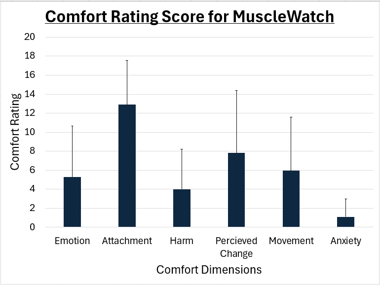
Figure 12. Participants’ comfort evaluation of the MuscleWatch.
Discussion
- Overall, the results of this validation confirmed that the proposed MuscleWatch prototype is highly feasible, with participants finding the device wearable and enjoying having their real-time muscle activation state data made available to them during Neuromuscular Training (NMT) warm-up exercises.
- As for the determination of the MuscleWatch’s effectiveness in contributing to the improvement of exercise fidelity, positive exercise fidelity improvements were observed but they were exercise-specific:
- When shown the MuscleWatch predictions, participants’ exercise fidelity for the Nordic Hamstring Curl NMT warm-up exercise improved. This could be attributed to the Nordic Hamstring Curl being a targeted hamstring-intensive exercise with low activation of other muscles in the proximity. These findings suggested that the MuscleWatch worked better with warm-up exercises that have consistent, repeatable motions that do not drastically differ from user to user.
- There was not sufficient evidence to identify a distinct pattern or correlation based solely on the Rate of Perceived Exertion (RPE) prediction accuracy results (Figure 10). Both the Nordic Hamstring Curl (Figure 10b.) and the Squat Jump (Figure 10c.) had a similar RPE difference between the ground truth and the MuscleWatch prediction.
- It is worthy to note that the ground truth of RPE measurements is a subjective measure provided by each participant.
- One major finding I observed is that the Nordic Hamstring Curl has the best prediction accuracy on exercise fidelity (Figure 9), which indicates the MuscleWatch preforms better on hamstring-focused NMT exercises where the motion is repeatable and consistent across all participants.
- Another major finding is that the extent of the machine learning (ML) model training has a direct impact on both the exercise fidelity and RPE prediction accuracies. As the MuscleWatch is solely trained on my own biomechanical hamstring data, the MuscleWatch exercise fidelity ML prediction accuracy fluctuates when worn by other participants depending on their own physiological factors.
- The effectiveness of the MuscleWatch is incomparable to previous research that were focused on exercise fidelity during NMT warm-ups and have relied on visual observations [11] and a IMU (Inertial Measurement Unit) device to determine the movement quality [9].
Limitations
- Design: The estimated sEMG signal amplitude data in time domain is the only feature used to represent the hamstring muscle activation. Although this approach is consistently used in all phases of this project, the acquired sEMG data may not fully characterize the intensity and duration of the hamstring muscle activation.
- Design: The Bluetooth transmission speed between the signal acquisition module (Arduino) and the microprocessor module (Raspberry Pi) is limited to 8 pairs of ADC values per second and thereby restricts the flow of data resulting in the potential loss of quality hamstring muscle activation data.
- Development: All three machine learning (ML) models were trained on my own physiological hamstring muscle activation data resulting in a biased model.
- Validation: Only one of the two groups of prospective users was included in this scope of the simulation. The ML models were only trained by a healthy researcher, and the ML models were only validated on healthy participants.
- Validation: The intent was not to stress or test one’s biomechanical strain limits for rehabilitation purposes. Hence, only Level 1, low-intensity Neuromuscular Training (NMT) warm-up exercises were performed by the participants.
- Validation: The total number of participants’ exercise repetitions performed by the 11 participants across all three NMT exercises was 569. The total number of exercise repetitions I completed in training, validating, and testing the machine learning models was 999. Both numbers indicate that this is a very small-scale validation study where the results may not represent the wider teenager population.
- Validation: In evaluating the MuscleWatch’s ML prediction accuracy on exercise fidelity, it was compared against my judgement of the participants’ execution of the NMT exercise repetitions. Although my assessment was formulated with the exercise fidelity criteria (Table 2) in mind, I do not have the expertise of a professionally trained physiotherapist to fully evaluate exercise fidelity.
- Validation: The NMT exercises performed were conducted indoors versus the typical warm-up exercise conditions in outdoor settings for the sprint-based sports.
Next Steps / Future Considerations
- Perform a comparative assessment of the MuscleWatch with conventional pre-gelled Ag/AgCl electrodes and with multichannel sEMG analysis functionalities to evaluate the validity and/or effectiveness of both the MuscleWatch’s sEMG acquisition module and the microprocessor receiving module.
- Upgrade the wireless transmission system from Bluetooth to wireless internet to improve the quality of the sEMG hamstring muscle activation data by allowing more sEMG ADC values to be processed and thereby resulting in better model prediction accuracies.
- Explore options, including but not limited to the use of Electronic Textile sensors [27], to improve the wearability level of the MuscleWatch, particularly in the comfort dimensions of “attachment”, "perceived change", and “movement”.
- Scale up the sample size and improve the training of the machine learning models by including trainers of diverse ages, genders, and hamstrings physiological state, including both healthy athletes and injured athletes undergoing rehabilitation. This way, it will improve the representativeness of the prospective user population and minimize model bias.
- Evaluate the performance of the MuscleWatch under the typical warm-up exercise conditions in outdoor settings for the sprint-based sports.
- Invite a professionally trained physiotherapist to observe and evaluate whether the model trainers and validation participants are performing repetitions of the NMT exercises with proper exercise fidelity adherence [9].
Conclusion
The findings of this project indicate that:
- It is feasible to produce a wearable MuscleWatch prototype for under $500 that serves as a non-invasive monitoring tool in providing real-time (i.e. smooth, continuous monitoring with greater than 50% accuracy) machine learning predictions on exercise fidelity adherence based on sEMG measurements of hamstring muscle activation.
- Validation participants consider the proof-of-concept MuscleWatch to be useful, comfortable with acceptable wearability, and effective in improving their exercise fidelity during Neuromuscular Training (NMT) warm-ups.
- The proposed MuscleWatch serves as a first step in exploring the relationship between improving teenagers’ exercise fidelity adherence to hamstring-targeted NMT warm-up exercises (using machine learning predictions) and the prevention of hamstring injuries.
Future Applications:
- The MuscleWatch can be used as a clinical rehabilitation tool in unsupervised settings for:
- prospective users who have been recently injured and are working on retraining their hamstring muscles through progressive loading during their rehabilitation exercise program; this approach gamifies their rehabilitation program while enhancing adherence to exercise fidelity; and
- prospective users who have a history of past injury and are trying to prevent re-injury upon return to play; The machine learning predictions provided by the MuscleWatch can help with improving exercise fidelity adherence in their NMT warm-up programs, thereby further reducing the risk of re-injury.
- Incorporating MuscleWatch as a real-time feedback tool to improve an athlete’s adherence to their NMT warm-up program allows recreational or community players:
- to develop consistent and effective habits in performing a prescribed NMT warm-up program,
- to achieve individualized warm-up goals in building their NMT capabilities, and
- to increase self-awareness of respecting their own biomechanical strain limits, which can ultimately prevent hamstring injuries.
- The machine learning capabilities of the MuscleWatch in providing real-time biomechanical feedback make it a very valuable tool for industrial athletes in an occupational environment for ergonomics monitoring and enhancements [28].
Citations
- Nakao, G., Kodesho, T., Yamagata, K., Watanabe, K., Ohsaki, Y., Katayose, M., & Taniguchi, K. (2024). Stress–strain relationship of individual hamstring muscles: A human cadaver study. Journal of the Mechanical Behavior of Biomedical Materials/Journal of Mechanical Behavior of Biomedical Materials, 153, 106473–106473. https://doi.org/10.1016/j.jmbbm.2024.106473
- Bramah, C., Mendiguchia, J., Dos’Santos, T., & Morin, J.-B. (2023). Exploring the Role of Sprint Biomechanics in Hamstring Strain Injuries: A Current Opinion on Existing Concepts and Evidence. Sports Medicine, 54(4), 783–793. https://doi.org/10.1007/s40279-023-01925-x
- O'Sullivan, L., Preszler, J., & Tanaka, M. (2022). Hamstring Injury Rehabilitation and Prevention in the Female Athlete. International journal of sports physical therapy, 17(6), 1184–1193. https://doi.org/10.26603/001c.38254
- Emery, C. A. (2023, June 6). Injury prevention research to reduce youth-sport related injuries. Open Access Government. https://doi.org/10.56367/OAG-039-10903
- Leonardelli, C., Howitt, S., & deGraauw, L. (2023). Canada-based sports chiropractors' attitudes, beliefs, and practical application of sport psychology in the injury rehabilitation process: a mixed-methods study. The Journal of the Canadian Chiropractic Association, 67(3), 226–245.
- Haraldsdottir, K., & Watson, A. M. (2021). Psychosocial Impacts of Sports-related Injuries in Adolescent Athletes. Current Sports Medicine Reports, 20(2), 104–108. https://doi.org/10.1249/jsr.0000000000000809
- Jeong, L., & Li, D. (2024). Psychological Well-Being From Sports Injuries in Adolescence: A Narrative Review. Cureus, 16(7), e64018. https://doi.org/10.7759/cureus.64018
- Black, A. M., Meeuwisse, D. W., Eliason, P. H., Hagel, B. E., & Emery, C. A. (2021). Sport participation and injury rates in high school students: A Canadian survey of 2029 adolescents. Journal of Safety Research, 78, 314–321. https://doi.org/10.1016/j.jsr.2021.06.008
- Benson, L. C., Räisänen, A. M., Sidhu, S. S., & Emery, C. A. (2022). Comparison of Measured and Observed Exercise Fidelity during a Neuromuscular Training Warm-Up. Biomechanics, 2(3), 361–373. https://doi.org/10.3390/biomechanics2030029
- Kalema, R. N., Schache, A. G., Williams, M. D., Heiderscheit, B., Siqueira Trajano, G., & Shield, A. J. (2021). Sprinting Biomechanics and Hamstring Injuries: Is There a Link? A Literature Review. Sports, 9(10), 141. https://doi.org/10.3390/sports9100141
- Perera, N. K. P., & Hägglund, M. (2020). We have the injury prevention exercise programme, but how well do youth follow it? Journal of Science and Medicine in Sport, 23(5), 463–468. https://doi.org/10.1016/j.jsams.2019.11.008
- Räisänen, Anu M.; van den Berg, Carla; Owoeye, Oluwatoyosi B.A.; McKay, Carly D.; Emery, Carolyn A. Running Every Time, Planking Sometimes: Youth Adherence to a Neuromuscular Training Program. Translational Journal of the ACSM 7(4):e000209, Fall 2022. | DOI: 10.1249/TJX.0000000000000209
- Hansen, J. T., Frank Henry Netter, Machado, C. A. G., Dragonfly Media Group, & Amsterdam, E. (2019). Netter’s anatomy coloring book. Elsevier.
- Raiser, S., Maier, J., Weppner, J., Negron, G., & Herman, D. (2017, February 28). Proximal and Mid-Hamstring Strain/Tendon Tear. PM&R KnowledgeNow. https://now.aapmr.org/proximal-and-mid-hamstring-straintendon-tear/
- Seçkin, A. Ç., Ateş, B., & Seçkin, M. (2023). Review on Wearable Technology in Sports: Concepts, Challenges and Opportunities. Applied Sciences, 13(18), 10399–10399. https://doi.org/10.3390/app131810399
- Wu, D., Tian, P., Zhang, S., Wang, Q., Yu, K., Wang, Y., Gao, Z., Huang, L., Li, X., Zhai, X., Tian, M., Huang, C., Zhang, H., & Zhang, J. (2024). A Surface Electromyography (sEMG) System Applied for Grip Force Monitoring. Sensors, 24(12), 3818–3818. https://doi.org/10.3390/s24123818
- Analog EMG Sensor by OYMotion SKU:SEN0240. (n.d.). Wiki.dfrobot.com. https://wiki.dfrobot.com/Analog_EMG_Sensor_by_OYMotion_SKU_SEN0240
- Milosevic, B., Benatti, S., & Farella, E. (2017). Design challenges for wearable EMG applications. Design, Automation & Test in Europe Conference & Exhibition (DATE), 2017. https://doi.org/10.23919/date.2017.7927217
- OYMotion, Inc. (2017). EMGFilters/EMGFilters.h at master · oymotion/EMGFilters. GitHub. https://github.com/oymotion/EMGFilters/blob/master/EMGFilters.h
- Hikaru Shimada. (2021, January 10). AI Arduino EMG Sensor !!!! Tensorflow on a Raspberry Pi !!! YouTube. https://www.youtube.com/watch?v=QjK82QfulCU&t=35s
- Paolo Perrotta. (2020). Programming machine learning: from coding to deep learning. Raleigh The Pragmatic Bookshelf.
- Clancy, E. A., Morin, E., Hajian, G., & Merletti, R. (2023). Tutorial. Surface electromyogram (sEMG) amplitude estimation: Best practices. Journal of Electromyography and Kinesiology, 72, 102807–102807. https://doi.org/10.1016/j.jelekin.2023.102807
- Cleveland Clinic. (2023, November 1). Rated Perceived Exertion (RPE) Scale. Cleveland Clinic. https://my.clevelandclinic.org/health/articles/17450-rated-perceived-exertion-rpe-scale
- Warburton, D., Jamnik, V., Bredin, S., Shephard, R., & Gledhill, N. (2022). The 2022 Physical Activity Readiness Questionnaire for Everyone (PAR-Q+) and electronic Physical Activity Readiness Medical Examination (ePARmed-X+): 2022 PAR-Q+. The Health & Fitness Journal of Canada, 15(1), 54–57. https://doi.org/10.14288/hfjc.v15i1.815
- Sport Injury Prevention Research Centre, Faculty of Kinesiology, University of Calgary. (2023, February 16). SPIRC-SHRED Injuries: All Sports. University of Calgary. https://ucalgary.ca/shred-injuries/all-sports
- Knight, J. F., Baber, C., Schwirtz, A., & Bristow, H. W. (2002). The comfort assessment of wearable computers. Proceedings. Sixth International Symposium on Wearable Computers. https://doi.org/10.1109/iswc.2002.1167220
- Amitrano, F., Coccia, A., Pagano, G., Biancardi, A., Tombolini, G., Marsico, V., & D’Addio, G. (2024). Measuring Surface Electromyography with Textile Electrodes in a Smart Leg Sleeve. Sensors, 24(9), 2763–2763. https://doi.org/10.3390/s24092763
- Marulli, A., Irvine, E., & Rojas, H. (2014). Functional rehabilitation – PM&R KnowledgeNow. Aapmr.org. https://now.aapmr.org/functional-rehabilitation/
- Slocum, A. (2008, February 1). FUNdaMENTALS of Design. Mit.edu. https://pergatory.mit.edu/resources/FUNdaMENTALS.html
Acknowledgement
I would like to thank these individuals who have supported me throughout this project:
- Dr. Karen Ballinger, MD FRCPC, Pediatrician at the University of Calgary Sports Medicine Centre for her invaluable medical advice on youth sports injuries and hamstring injury prevention strategies.
- Mr. John Schipilow, PT, MScPT, MScBME, BScBiomech of the University of Calgary Sports Medicine Centre for offering constructive feedback on design improvements, sharing his expertise on surface electromyography clinical applications in the context of injury rehabilitation, and advising me on effective technical communications.
- Dr. Cody Dunne, Primary Investigator, Department of Community Health Sciences at the University of Calgary, for letting me observe how he conducted his simulation study and learn about the process of obtaining consent to participate in a research study.
- Mr. Ross Thompson, Science Fair Club Teacher Sponsor at Colonel Irvine School for safeguarding all the signed consent forms for my project and sharing his passion and expertise in science fair participation.
- My father for funding this project and sharing with me his MS Excel and PDF editing expertise.
- My mother who plays the role of a demanding yet supportive business client, setting firm project execution requirements and often reminding me to learn from the experts in each field in the pursuit of excellence. I appreciate mom taking the time to review all my drafts and ask tons of questions to help me improve the clarity of my written communication. Thank you mom for your never-ending encouragement and faith in me!
- My sister whom I can always count on to be there for me no matter where she is. She is my role model, my inspiration and best of all, she always believes in me!
And above all, a huge shout out to the Colonel Irvine School student athletes who participated in my validation study of this project. Getting to spend time with you has already made this lengthy project truly worthwhile.