The NEUROWATCH: A Neural Electrical Understanding and Response Optimizer that uses a BCI system to treat Alzheimer's disease.
Aaryan Praveen
STEM Innovation Academy High School
Grade 10
Presentation
No video provided
Problem
1.0 ABSTRACT
Alzheimer's disease affects million of people every year, often being the main cause of dementia in older individuals. In addition to dementia, victims of the disease experience cognitive decline, behavioral and personality changes, and also things like infections and seizures due to the lack of control of the biological processes in their body. Eventually patients of Alzheimer's become unrecognizable from the person they once were, severing connections, interaction and memory of their loved ones. Current treatments help slow down the overall effects of Alzheimer's, targeting the biochemical progression of the disease, yet do not adjust to variability, modify in real time, and do not specifically influence certain behaviors or personality. My project attempts to improve the lives of Alzheimer's patients, filling in the gaps that certain treatments have. Throughout my project, I explore the concept of developing a non-invasive brain computer interface(BCI) that interprets neural activity from the brain and responds with adaptive electrical signals to support neural electrical communication within the body. The NEUROWATCH analyses data from an open source electroencephalogram(EEG) platform and differentiates between normal neural activity, and one from an Alzheimer's patient. This is achieved through the use of a micro controller(Raspberry Pi/Micro bit) and Python script that looks for variation within the EEG signal and classifies it as either stable or unstable. If a signal is classified as unstable, commands are sent from the micro controller to a PWM driver that generates and shapes electrical responses depending on the type of variation. These signals are then sent to an output or load, continuing the response until stability is achieved. My project evaluates a potential solution to how neural stability can be maintained within Alzheimer's patients, allowing for more focused brain to body coordination when trying to complete tasks.
1.2 INTRODUCTION
Every year millions of people are diagnosed with life-threatening diseases that affect their daily lives, prevents them from doing certain activities, and forces them to follow strict treatments in order recover. Yet some diseases are not so simple, as they are fatal, incurable, and you never see it coming. One of the biggest examples is Alzheimer's disease. Often the cause of dementia, Alzheimer's disease interferes with the ability to complete daily tasks and comprehend information such as remembering a loved one’s name. In 2021, 57 million people worldwide were diagnosed with Alzheimer's with nearly 10 million new cases reported every year. Though there are biochemical treatments to Alzheimer's, they are often not readily available in certain locations due to geographical, and diagnostic barriers. With limited options to counter such a disease and its difficulty in even diagnosing or recognizing it, most people ignore its effects and the associate it with symptoms of old age.
1.3 PROBLEM
Problem: Can the effects of Alzheimer’s disease be countered or improved upon through the use of a low cost brain computer interface(BCI) system? Hypothesis/Predicted Outcome: If you use current technologies such as a brain computer interface(BCI) to process information in the brain, then it could act as alternative solution to treat or counter Alzheimer’s disease because research has already proven that it is possible to interpret and analyze information perceived through the brain using artificial processes.
Objective/purpose: To develop a new tool or product that interprets information sent by the brain and responds accordingly to the body, assisting Alzheimer’s patients to complete tasks and live normal lives.
Method
2.0 - Goal
Due to how life threatening Alzheimer's disease is, many treatments have been created, most of them being drug based treatment. Currently the most common treatment is donepezil, a cholinesterase inhibitor that works by increasing acetylcholine levels. Acetylcholine is basically a neural transmitter that acts as a chemical messenger. The main problem with these drug based treatments is that they have side effects, only temporarily slow down the effects of the disease and are only effective if the Alzheimer patient is diagnosed at an early stage. Pre-existing damage can not be repaired and lost cognitive skills can not be regained. In my project, I am attempting to develop a new product for Alzheimer's patients that interprets information by analyzing electroencephalogram(EEG) data sent to the brain and then responds with corresponding actions sent through the rest of the body. Through the controlled, neural-electrical signals sent out from the proposed device, Alzheimer's patients could lead normal lives and have reduced setbacks associated with Alzheimer's disease.
2.1- What is Alzheimer's Disease?
Alzheimer's disease is a progressive brain disorder that disrupts memory, judgement, thinking skills, and and the understanding of information. The disease slowly gets worse, over time, eventually leading to the difficulty performing common tasks that are usually second nature. The lack of interpretation and understanding of information results mood swings, changes in personality or behavior, asking the same question multiple times, forgetting where common household object are, forgetting certain names or words and even confusing or questioning reality. Eventually when the disease develops to one of its final states, the patient becomes completely reliant on someone else or external support in order to survive as their cognitive skills and control of their body has become essentially been destroyed. As of 2026, over 7.2 million Americans over the age of 65 have been diagnosed with Alzheimer's, between 700 000-800 000 Canadians have been diagnosed with a form of dementia, and over 55 million people globally are said to have dementia. Scientists predict that this number will sky rocket to 78 million people by the year 2030. In addition to this, in high income countries, only 20-50% dementia cases are recognized and diagnosed while in low income countries such as India, 90% of cases are left undiagnosed. Considering that dementia is just an umbrella term for the much more serious Alzheimer's disease to fall under, it could mean that even more people are left undiagnosed.
2.1.1 - What causes Alzheimer's Disease?
Most of the time, a patient gets Alzheimer's disease through multiple ways such as genetics, age, lifestyle, environment, medical conditions etc. In fact, it said a patient can have had Alzheimer's 20 years before they have any noticeable symptoms, showing how deadly the disease really is.
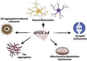
Genetics: As a person gain genes from their parents, a parent with a variation in the genes can cause the development of Alzheimer's disease in their offspring. Yet in most cases, Alzheimer's is not developed due to one specific gene but a rather a combination of variations mixed with lifestyle and environmental factors as well. A common gene that leads to Alzheimer's when it has variation is apolipoprotein E (APOE). The APOE gene is involved in making a protein that carries fats and cholesterol into your blood stream. But if this protein becomes dysfunctional, it promotes the creation of amyloid-beta plaques and tau tangles, which ultimately leads to the destruction of nerve cells. APOE comes in multiple forms known as alleles. A child receives 2 alleles from each parent. If the APOE ε4 allele is received, the child has a higher risk in developing Alzheimer's later in life.
Other variations with genes that lead to Alzheimer's include:
- Amyloid precursor protein (APP) on the 21st chromosome
- Presenilin 1 (PSEN1) on the 14th chromosome
- Presenilin 2 (PSEN2) on 1st chromosome
If a parent has variation within any of these three genes, their offspring has a 50/50 chance of inheriting that variation and therefore increasing the risk of developing Alzheimer's disease.
Lifestyle: Scientists have found that certain lifestyle factors may contribute or associate with the development of Alzheimer's disease. Certain medical conditions such as hearing loss, depression, concussions or brain injuries, and mild cognitive impairment may increase the risk of Alzheimer's. In addition to that, lifestyle choices that also contribute to the development of the disease are:
- Social Isolation
- Smoking
- Over-Consumption of Alcohol
- Unhealthy diet
- Lack of sleep
- Being physically inactive
- And unmanaged chronic health issues such as blood pressure
Those these factor do not immediately develop into Alzheimer's, the affects it has on your biological systems over time is what eventually results in the development of the disease.
Age: As a person's age increases, their biological processes and systems become weaker, making the brain and body more vulnerable to the disease. With the immune system being much slower and less efficient in removing harmful substances from your body, plaques and tangles only increase, which allows the progression of the disease. 1 in 20 people over the age of 65 have Alzheimer's disease while 1 in 4 people over the age of 85 have the disease.
Damage starts at the hippocampus, the part of the brain that deals with memories. From there it slowly spreads throughout the brain, taking over the brain and killing its cells. Eventually the part of the brain that control decision making and judgement is take over, resulting in your body becoming physically uncappable of living on its own.
2.1.2 - How does Alzheimer's affect the brain?
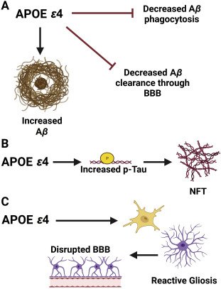
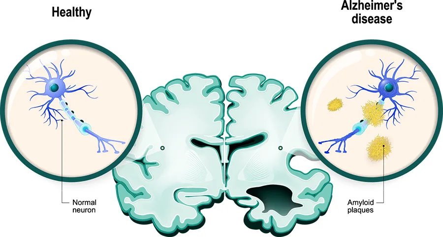
Alzheimer's is caused by the abnormal buildup of proteins in the brain that lead to amyloid plaques and neurofibrillary tangles in the brain. Normally within the cell membrane of a neuron, there is a protein called Amyloid Precursor Protein (APP). This protein play in important role in neural growth, repairing neurons, maturation during brain development and cell fate specification. Once this protein is used, it is broken down by enzymes called alpha secretase and gamma secretase, creating a peptide that is soluble and dissolves away. Yet if another protein called beta secretase slices APP with gamma secretase instead of alpha secretase, it creates an insoluble monomer called amyloid beta. These monomers stick and bond together outside neurons, creating this clump called beta-amyloid plaques. These plaques may buildup between neurons, disrupting neural, electrical signals that prevent brains cells from relaying information. This can result in the loss of certain brain functions like memory or decision making. In addition, the beta-amyloid plaques creates an immune response that results in surrounding inflammation, in which damages the neurons. Amyloid plaques will also gather around blood vessels in the brain, called amyloid angiopathy. This weakens the structure of the vessels, leading to hemorrhage and blood loss.
The other factor for Alzheimer's is neurofibrillary tangles within the neuron. Neurons, being any other type of cell are held together through their cytoskeleton, which consist of microtubules. The microtubules transfer nutrients around the cell. They are held together with the tau protein, but when the beta-amyloid plaque builds up outside the neuron, it initiates the activation of kinase, a enzyme that helps transfer phosphate groups to tau proteins. This modifies the shape of the tau proteins and causes them them to tangle together, no longer supporting the structure of the microtubules. With breaks between microtubules, signals and nutrients can not be transferred and may result in apoptosis.
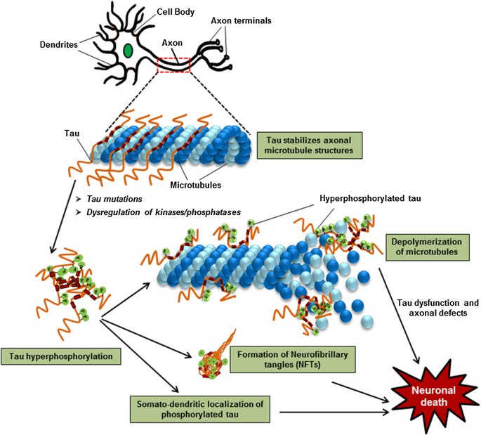
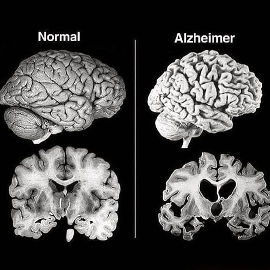
With the death of neurons and brain cells, the brain shrinks. The edges around the brain called gyri become narrower while the small gaps between parts of the brain become wider. The fluid filled sections of the brain called ventricles also become larger as well.
Key Words:
- Monomers- Molecules that react with each other to create chain-like structures called polymers
- Hemorrhage-The rupture of a vessel
- Amyloid Precursor Protein- A protein responsible for neural growth and repair
- Beta-amyloid plaques- Clumps of amyloid protein that were sliced by gamma and beta secretase, resulting in an insoluble monomer that disturbs neural communication
- Apoptosis- Programmed cell death where the immune system destroys damaged or old cells.
The images below shows the difference between a healthy brain and a Alzheimer's brain. As plaques increase, more neurons and brain cells die, leading to the shrinkage and cognitive decline of the brain.

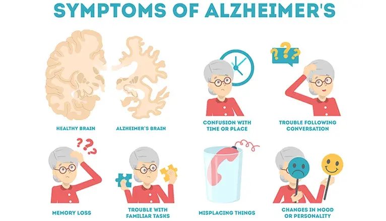
2.1.3 - Side Effects of Alzheimer's
Alzheimer’s has multiple side effects that continuously gets worse as the the disease progresses. These include:
- Dementia/forgetting well known or basic knowledge.
- Making improper judgement(Ex. Dressing appropriately depending on the occasion).
- Trouble communicating or naming certain objects.
- Misplacing or grabbing the wrong things.
- Withdraw from work or social activities.
- Make unusual decisions that are not normal for the person.
- Confusion interpreting time or places.
- Depression.
- Imagining things.
- Questions reality.
-
Loose motor function.

Living with such conditions makes it impossible to live a comfortable life due to the difficulty to understand and process information. Not only does this affect the patient so greatly, but it also affects their family and friends as Alzheimer's creates a communication gap between them, making it hard to rely information and create connection.
2.1.4 - Current Alzheimer's Treatments
Current Alzheimer's treatments are all drug based in which aims to either eliminate beta-amyloid plaques or increasing cell to cell communication. The following are examples of current treatments:
- Cholinesterase inhibitors: These work by boosting the levels of acetylcholine, a neurotransmitter involved in memory, attention, and neural communication. In Alzheimer's acetylcholine usually become depleted so the increase in these chemical messengers help create temporary improvement in symptoms. Pros: -Temporarily improves cognitive strength -Makes symptoms less noticeable -Affordable -Easy to prescribe Cons: -Diarrhea, nausea, loss of appetite and trouble with sleep -Does not stop or slow down the progression of disease
- Donanemab: Works by eliminating beta-amyloid plaques using immune cells. Pros: -Can alter the progression of the disease -Slows cognitive decline in early Alzheimer's Cons: -Expensive -High maintenance -Risk of ARIA (amyloid-related imaging abnormalities) as well as interal swelling and bleeding in the brain -Ineffective in severe Alzheimer's
- Memantine (Namenda): In Alzheimer's NMDA receptors are overstimulated, leading to calcium overload, oxidative stress and neuron death. This drug blocks the activation of NMDA receptors which stabilizes neural networks and signaling. Pros: - More tolerable than Cholinesterase inhibitors as they are consumed orally as either a tablet or liquid. -Can be used along with other treatments -Slightly slows cognitive decline Cons: - Ineffective in early Alzheimer's if used alone -Cause confusion, headaches, dizziness, and high blood pressure -Does not remove tangle or plaques.
Other treatments also include non-drug based cognitive and behavioral therapies aimed to improve mental stimulation and memory but these treatments do not change any affect Alzheimer's has had on a paitents brain.
2.2 - Neural Electrical Understanding
2.2.1 - Neural Electrical Impulses
Neural Electrical impulses are the signals your brain send throughout your body to complete certain action. Everything from walking to watching a movie makes your body send signals to the brain on collected information. The brain interprets this information and sends signals back to each part of the body so it can respond correctly. Without such signals, the body can not perform correct functions. Things like digestion would not happen, causing waste intoxication in your body. The nervous system and neurons play such an important role in having a functional body that without it, you would not be able to survive. There are two nervous systems in your body, the central nervous system and the peripheral nervous system:
Central nervous system: Is made up of your brain and spinal chord. Your brain processes signals from your nerves to then respond with signals corelated with thought, movement, and feeling.
Peripheral nervous system: Is made up of a network of neurons around your body that all branch out from the spinal chord. The peripheral nervous system relays information sent by the brain to the rest of the body, ensuring that signals for biological processes reach their location.

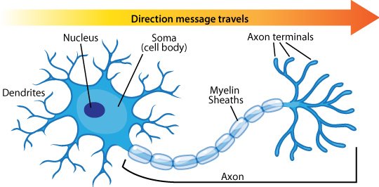
Each neuron consists of a cell body(soma), an axon that sends signals to muscles other neurons and muscles and dendrites that interpret signals. If stimulated enough, the neuron generates electrical impulse called as action potential that trigger the release of neurotransmitters from synapses.
When these chemical messages are all sent my a network of neurons to the brain, the brain takes in that information to respond with control of states and systems in your body.
2.2.2 - Patient Monitoring
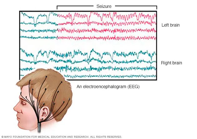
As millions of neurons produce signals, these signals can be measured through the scalp using electroencephalography(EEG). An EEG works by having electrodes measure voltage fluctuations in your brain. This data can be used to understand the activity in your brain, see patterns, and convert to control signals for BCl systems. Using an EEG allows for precise data as it filters out sound from muscles and other parts of your body, only scanning for information from neurons.
2.2.3 - Brain Computer Interface
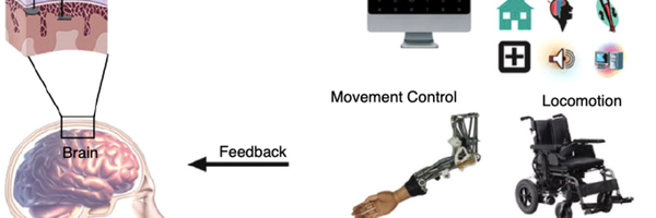
A brain computer interface(BCI) is a systems that acts as the direct line of communication between brain signals and a device such as a computer or robotic part. This system analyzes neural signals, and converts them into controls that be used to operate the device. This can allow you to regain muscle movement or motor function without the need of implants or surgery.
2.2.4 - Signal Interpretation
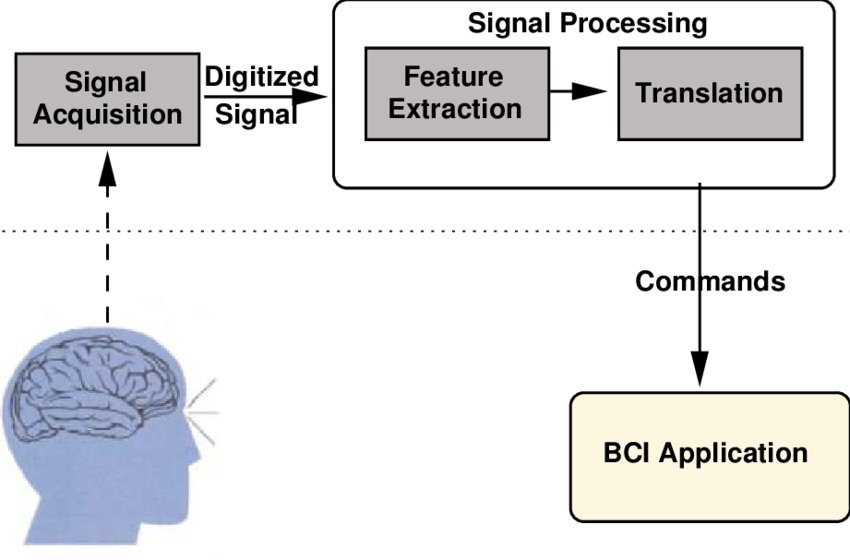
A BCI system requires multiple components to work but in simple terms these components are:
Signal Acquisition: The process of recording and collecting data on brain signals.
Signal Processing: Amplifying raw signals, with external noises and distractions being filtered out.
Extraction and Classification: Data is analyzed and categorized based on mental states or intentions.
Output: Signals are converted to usable control for an external device.
2.2.4.1 - Electroencephalogram(EEG) signals
EEG signals refer to the recording of the brain's spontaneous electrical activity over a period of time, recorded from multiple electrodes placed on the scalp. It can be used to measure and interpret neural activity in the brain and understand neural states.
2.2.4.2 - Signal Conversion
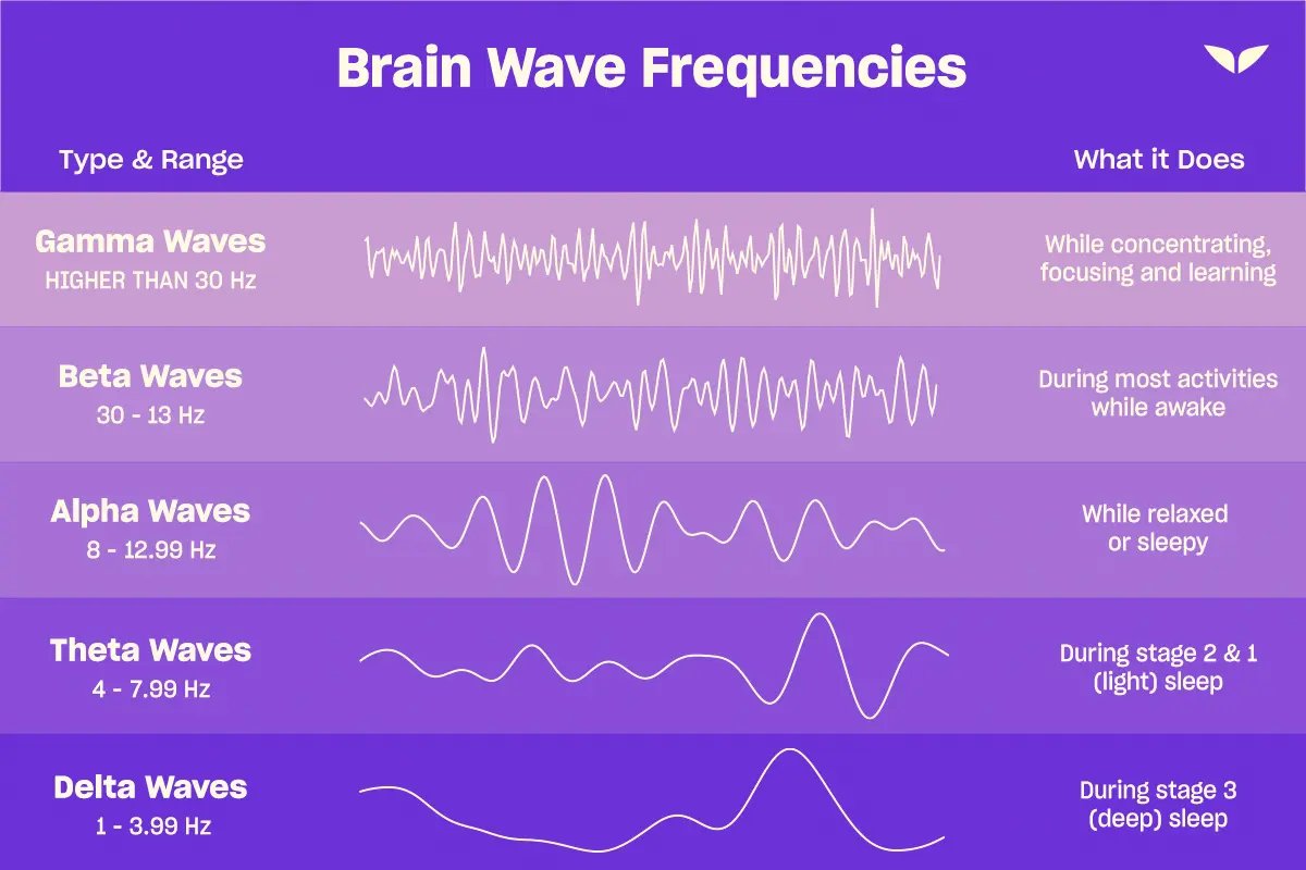
Electrodes placed on the scalp measure electrical activity using a process called electroencephalography. As signals are passed among neuron, small voltages are created and measured. As these voltages are really weak, they are amplified using a bio-potential amplifier, making each signal more prominent. From there, signals are filtered down to remove high frequency noise and any electrical interferences. The frequencies can fall under 5 different categories listed below: Depending on the type of siganl wanted, different filter can be put in place.
| Brain Wave | Frequency |
|---|---|
| Delta | 0.5–4 Hz |
| Theta | 4–8 Hz |
| Alpha | 8–13 Hz |
| Beta | 13–30 Hz |
| Gamma | 30–100 Hz |
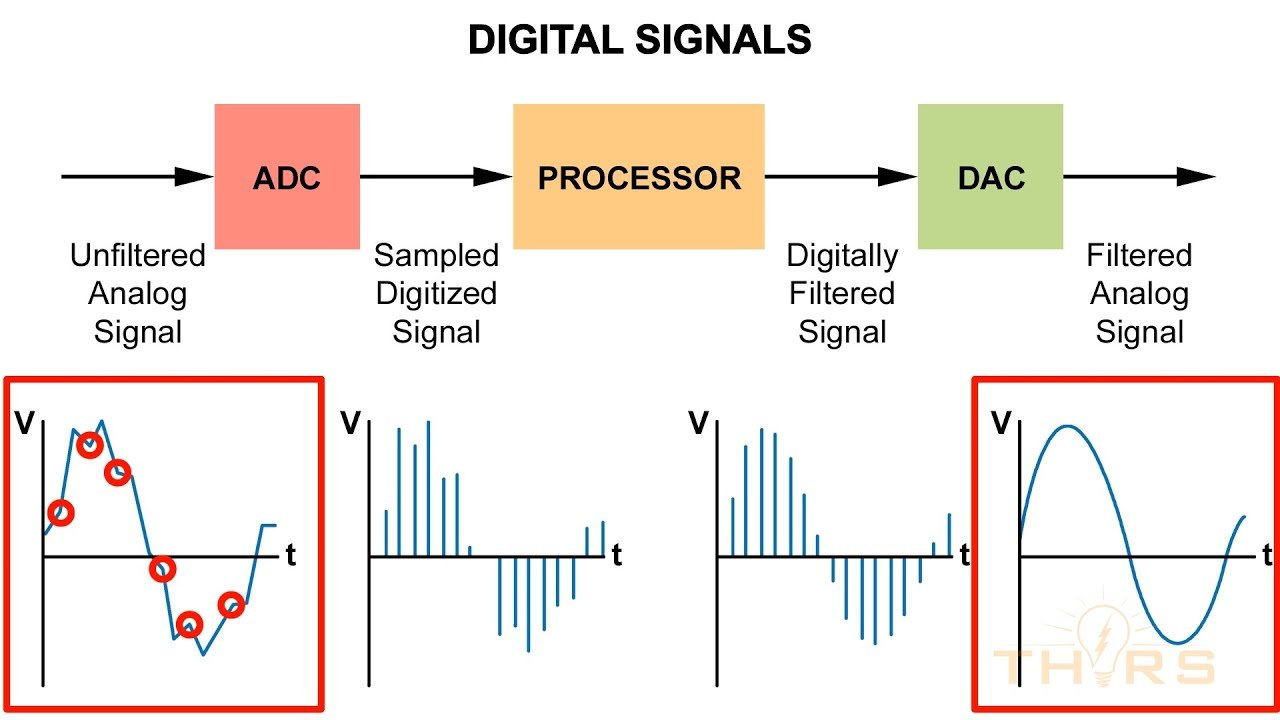
Once a brain computer interface extracts the raw signals, it converts it from analog to digital, changing the value from Hertz to voltage over time.
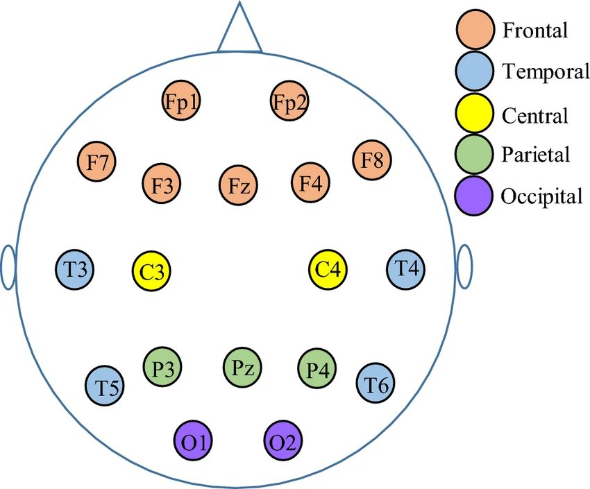
2.2.4.3 - EEG Translation
EEG signals are separated into channels that represent which electrode the data is being collected from. Odd numbers correspond to the left half of the brain while even numbers correspond to the right side of the brain. The letter represent the location of the electrode. For example channel T4 represent the right temporal. Anything ending in z refers to the midline of the brain. Below shows the abbreviation and name of each electrode location.
|
| Abbreviation | Name |
|---|---|
| N | Naison |
| Fp | Frontal polar |
| AF | Antero-Frontal(Anterior Frontal) |
| F | Frontal |
| T | Temporal |
| FT | Fronto-Temporal |
| FC | Fronto-Central |
| C | Central |
| TP | Temporo-Parietal |
| CP | Centro-Parietal |
| P | Parietal |
| PO | Parieto-Occipital |
| O | Occipital |
| I | Inion |
Pg Nasopharyngeal
Below shows the location for electrode placement:
When trying to understand what EEG signals translate to when considering the activity that is being done, you have to look at the frequencies, timing and amplitude of the signal. Depending on the type of frequency, the signals are categorized into different neural states. The exact activity can not be read but if you read the type of signals being sent and the location they are being sent from, you can create a rough picture of what the person is doing. Combinations of certain signals also may further clarify the task that is trying to be achieved. When considering brain regions, the occipital lobe correlates to vision processing, the motor cortex corresponds to movement planning and the and the frontal lobal corresponds to decision making. In addition the more waves there are per time base, the more electrical activity hat is taking place. For example something like sleeping and playing a video game are going to have difference concentrations of brain waves as one requires more neural activity in order to function. In Alzheimer's there may be certain abnormalities in brain waves such as have too little brain waves than would be consider usual when trying to to complete that task.
Below is an image of the types of waves associated with a type of neural state:
Safety and Goal
Create a system using the research on BCI systems that interprets neural signals using the EEG, processes it, and responds by completing a series of actions that corresponds with the signals perceived. I want to make sure that this device not only functions without error but does not damage the neural network of consumers, is not invasive(implanted into the the skull), is built through low cost materials, does not pose as difficult to use and maintain, and allows you to regain motor and cognitive control in your body.
3.0 - Developing the Brain Computer Interface
3.1 - Procedure
- Place EEG on the surface where data is being recorded.
- Connect the signal amplifier to the EEG through it’s inputs
- Attach the power source indirectly to reduce electrical noise.
- Open EEG software(PhysioNet) and record date. Data should be represented in a graph where each spike corresponds to specific signals.
- Filter the EEG data to record the best results.
- Export data and program microcontroller to conduct a command based on the spike seen in the graph.
- Complete tests multiple times to record patterns.
- Use data to have the EEG signals control motor output as well as program a user interface that runs by completing a certain function based on the signal detected.
* I won't be conducting live EEG testing but rather using an open source EEG platform for the data required to run the BCI.
3.1.1 - Materials
- Dry EEG electrodes
- A low cost amplifier chip(AD8232)
- Microcontroller such as an Arduino or Raspberry Pi
- Output source(A computer)
- Loads(Servos, dc motors, LEDs)
- A biopotential amplifier
- Analog to Digital convertor(ADC)
- 3D printed frame to house the components of the BCI system.
*As I am receiving my EEG signal from PhysioNet, dry EEG electrodes and a biopotential amplifier will not be necessary. Also certain components may be changed or not be used as certain materials are not mandatory in order for the system to function.
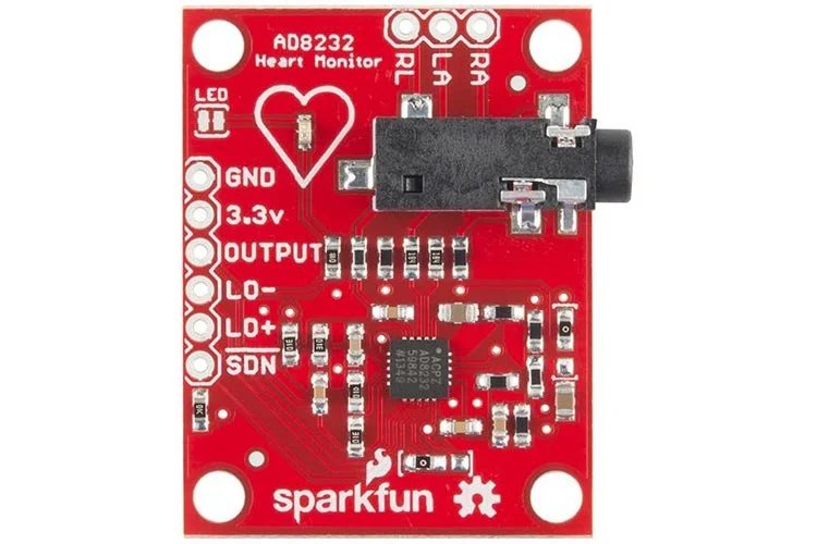 Amplifier chip-Strengthens EEG voltage and signal accuracy
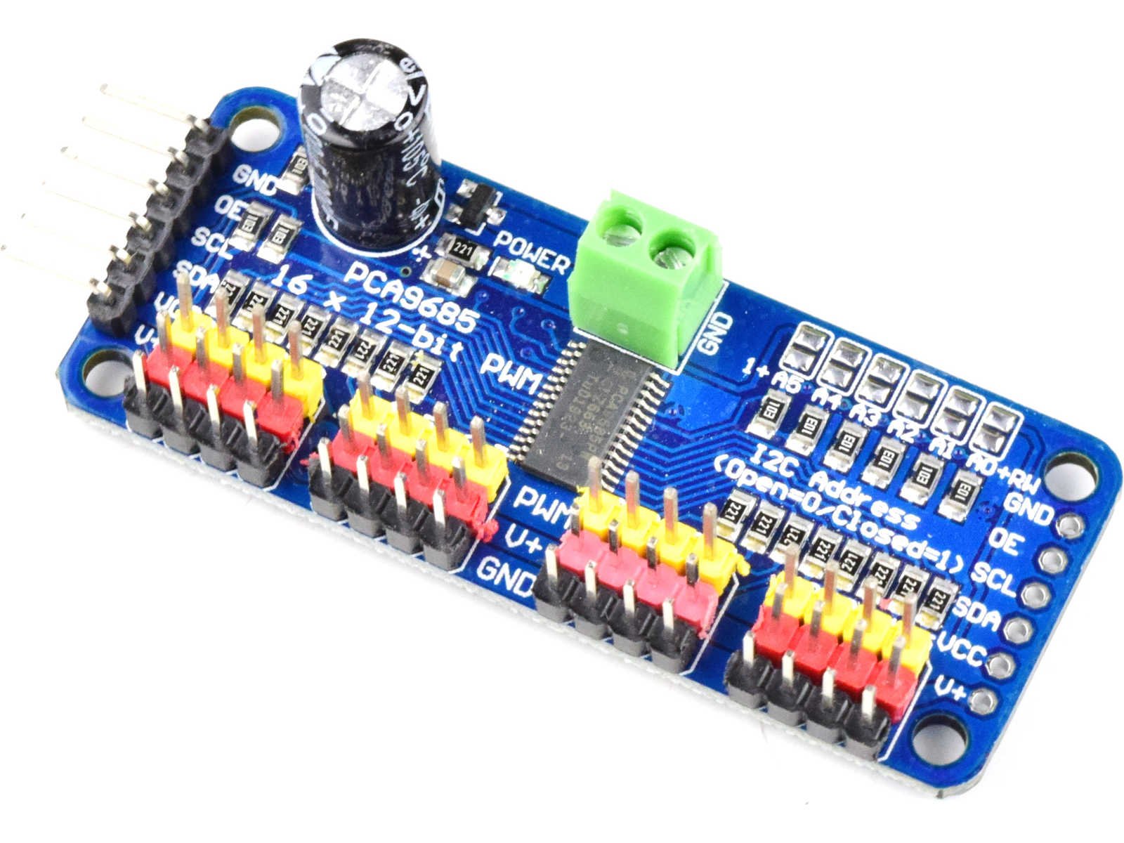 PWM driver-Controls the output through calculated electrical pulses
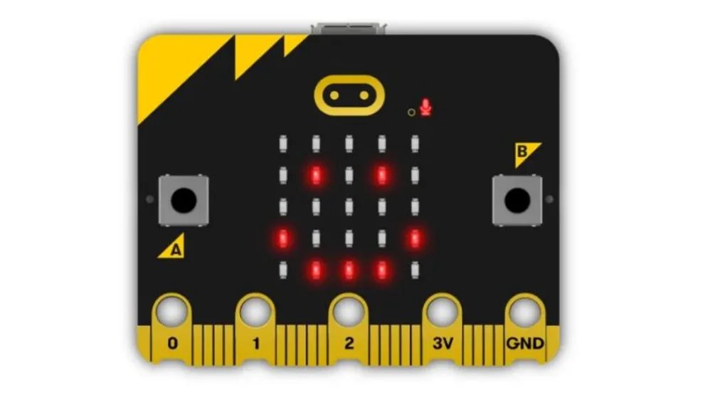 Micro bit-delivers feedback to ensure the electrical response has been received
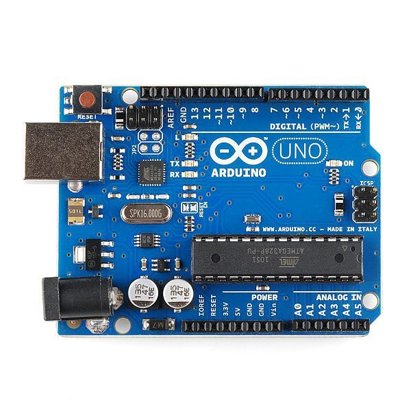 Arduino Uno-Controls hardware responses, taking in commands from the raspberry pi to initiate an electrical response
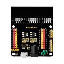 Micro bit sensor shield-Controls output and allow for additional sensors
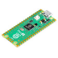 Raspberry Pico- Converts the analog data into digital data in order for the interface to interpret EEG signals.
In my project I will be analyzing the brain waves associated a type of neural state. If any abnormalities are detected, my machine will have an alert and will initiate an electrical response. I will demonstrate this by using a raspberry pi 5 microcontroller that will analyze the EEG data and while send a signal to an Arduino if instability is detected. Depending on the difference between the abnormal signal and a normal signal, my system will deliver a neural electrical signal related to completing that task. That signal is sent to a load which represents the neuroelectric response that would be sent into to the body to support signal communication. This signal will be be sent at a low frequency to prevent any damage but continuously be sent until the neural state of the brain stabilizes. In a real life scenario, the response would be transcranial electron stimulation, which are small electrical signals sent through electrodes placed on the scalp. Originally I thought the response could be initiated through transcranial magnetic simulation(TMS) because of its stronger electrical stimulation. Yet I switched to the transcranial electron stimulation response because TMS is expensive, large and heavy, requires a large power source, and not generally potable or wearable as they are not intended for daily use.

Analysis
4.0 - Analysis of Data
This project aimed to see whether electroencephalography (EEG) signals sent from the brain could effectively be analyzed to detect variation within the neural electrical state of the person. Pre-existing EEG data on specific neural states were taken from a public dataset and processed using python. By plotting the EEG signals, I could visualize fluctuations and rhythmic oscillations. Each oscillation represented a different frequency of brain wave.
Sample dataset captured for experimental runs
-Baseline, eyes open
-Baseline, eyes closed
 Using the python script, process the data from “EDF” (European Data Format) files to perform
-Signal filtering
-Alpha Power Calculation
-Threshold detection
Using the python script, process the data from “EDF” (European Data Format) files to perform
-Signal filtering
-Alpha Power Calculation
-Threshold detection

Using Python script, the dataset was classified for High, Medium and Low engagement. This was done by comparing alpha power of the signal to a median threshold

4.1 - Sources of Error and Success
Success: In this project I had many success all fueled through the repeated process of failing and iterating. These success include:
-Designing a new solution to Alzheimer's disease that supports neural electrical communication, allowing for patients to comprehend and relay information easier. This would result in a much slower cognitive decline, granting patients more control, stability, and time, even while having Alzheimer's disease
-Designing not only an effective design but a low cost one that can be used in low income countries, allowing for faster diagnosis of Alzheimer's disease and support of neural communication.
-Developing a software that properly differentiates between stable and unstable neural electrical frequencies, almost immediately finding variation and taking action. Not only does this system ensure that signals generated to the brain reach the rest of the body but it also assists in analyzing information sent from the body, back to the brain.
Sources of Error: I experienced many setbacks when it came to this project. The first setback was just not having an idea of what to do at the start of the project. My lack of experience when it came to neurological understanding made it difficult to progress in the project, especially when trying to figure out how to read EEG and how I could make a interface that could analyze them. This was eventually overcome through the many online sources, lessons, tutorials, and after reviewing with professionals and knowledgeable individuals. The second source of error was when developing the interface to detect neural states and find abnormalities. This again took a long time due to my great lack of experience. This required repeated trial and error before I was able to find somewhat of consistency between different EEG signals of the same neural state. Another error was on how to use some of the parts as I needed. I had never used a raspberry pi before. This was not a major setback as it was easily overcome through some assistance but it wasted a lot of my time and was often frustrating to use. The fourth setback is not necessarily that problematic but originally I was going to collect EEG data from a live EEG source but it completely skipped my mind that recording EEG data from the brain might be considered as human testing. For this reason I decided not to conduct any type of live EEG data collection and stuck with the open source platform. But because my system is scanning based on pre-existing data, there may be slight differences when conducting the experiment on a real human. The fifth source of error was that though my interface was efficient and worked properly, it was a relatively basic visualization of EEG data. With more precise data, neural electrical interpretation and response can become more advanced, being more similar to that of clinically used BCI. Finally the biggest setback was when developing my response to the abnormality detected from the brain wave. Even now, the response still is not always accurate towards the neural state. I have still not been able to identify the problem within my code that causes this inaccuracy but hopefully before the time of the fair, this issue can be resolved and I will be able to fully complete my project.
Conclusion
5.0 - Conclusion
Alzheimer's disease can have a major affect on people's lives, leading to dementia, the loss of motor control, confusion, difficult interpreting information, and etc. As of 2025, the disease has affected 772,000 Canadians with 414 new cases being reported everyday. 60-70% of all cases of dementia in Canada is caused by Alzheimer's, proving how many people have to live with such a condition everyday. With my invention, neural electrical communication can be interpreted properly and a correct corresponding electrical stimulation can be sent to neural networks, assisting with communication. The more concentrated signaling and control of biological processes can allow for more focused immune responses to neurofibrillary entanglement and beta-amyloid plaques. My proposed design can assist and even potentially challenge current drug based treatment to Alzheimer's, all with no health-opposing side affects. This low cost invention allows for faster diagnosis and slower cognitive decline. Not only could my project improve millions of lives but also bring families back together.
Citations
6.0 - Bibliography(APA)
Alzheimer’s Association. (2026, March 3). What is Alzheimer’s? https://www.alz.org/alzheimers-dementia/what-is-alzheimers
Alzheimer’s Association. (2026, March 3). Alzheimer’s disease causes and risk factors. https://www.alz.org/alzheimers-dementia/what-is-alzheimers/causes-and-risk-factors
Apex Hospitals. (2026, March 3). Understanding Alzheimer’s disease: A comprehensive guide for caregivers and loved ones. https://www.apexhospitals.com/blogs-articles/understanding-alzheimers-disease-a-comprehensive-guide-for-caregivers-and-loved-ones
Apex Hospitals. (2026, March 3). Understanding Alzheimer’s disease: A comprehensive guide. https://apexhospitals.com/blogs/understanding-alzheimers-disease-a-comprehensive-guide-for-caregivers-and-loved-ones-wbgjyd1
Arizona State University. (2026, March 3). Neuron anatomy. https://askabiologist.asu.edu/neuron-anatomy
Backyard Brains. (2026, March 3). Spike Recorder. https://backyardbrains.com/products/spikerecorder
BNCI Horizon 2020. (2026, March 3). Basics of brain-computer interfaces. https://bnci-horizon-2020.eu/about/basics
Canada.ca. (2026, March 3). Dementia highlights from the Canadian chronic disease surveillance system. https://www.canada.ca/en/public-
Cleveland Clinic. (2026, March 3). Nervous system. https://my.clevelandclinic.org/health/body/21202-nervous-system
Colwell, B. D. (2026, March 3). An introduction to the neuroscience of brain-computer interfaces. https://briandcolwell.com/an-introduction-to-the-neuroscience-of-brain-computer-interfaces-bcis
Cumming School of Medicine. (2026, March 3). What is a brain-computer interface? https://cumming.ucalgary.ca/research/pediatric-bci/bci-program/what-bci
Dorosti, S. (2026, March 3). EEG dataset. https://github.com/sarshardorosti/EEG_DataSet
Fiveable. (2026, March 3). Neuroscience: Brain-computer interfaces. https://fiveable.me/neuroscience/unit-13
Health Research Institute. (2026, March 3). EEG brain function measurement. https://www.healthresearch.org/eeg-brain-function-measurement
Hopkins Medicine. (2026, March 3). Electromyography (EMG). https://www.hopkinsmedicine.org/health/treatment-tests-and-therapies/electromyography-emg
International Alzheimer’s Disease. (2026, March 3). Dementia statistics. https://www.alzint.org/about/dementia-facts-figures/dementia-statistics
Krol, L. R. (2026, March 3). SEREEGA EEG simulation toolbox. https://github.com/lrkrol/SEREEGA
Mayo Clinic. (2026, March 3). Alzheimer’s disease symptoms and causes. https://www.mayoclinic.org/diseases-conditions/alzheimers-disease/symptoms-causes/syc-20350447
MedPage Today. (2026, March 3). Medical research news. https://www.medpagetoday.com/hematologyoncology/breastcancer/110123
National Institute on Aging. (2026, March 3). What is Alzheimer’s disease? https://www.nia.nih.gov/health/alzheimers-and-dementia/what-alzheimers-disease
National Institute on Aging. (2026, March 3). Alzheimer’s causes and risk factors. https://www.nia.nih.gov/health/alzheimers-causes-and-risk-factors/what-causes-alzheimers-disease
National Institute on Aging. (2026, March 3). Thinking about your risk for Alzheimer’s disease. https://www.nia.nih.gov/health/alzheimers-causes-and-risk-factors/thinking-about-your-risk-alzheimers-disease-five
National Institute of Neurological Disorders and Stroke. (2026, March 3). Brain basics: The life and death of a neuron. https://www.ninds.nih.gov/health-information/public-education/brain-basics/brain-basics-life-and-death-neuron
OHSU. (2026, March 3). Brain-computer interface systems. https://www.ohsu.edu/reknew/brain-computer-interface-systems
Parralab. (2026, March 3). NY head model. https://www.parralab.org/nyhead
Physiopedia. (2026, March 3). Introduction to neurophysiology. https://www.physio-pedia.com/Introduction_to_Neurophysiology
PubMed. (2026, March 3). Amyloid precursor protein and Alzheimer’s disease research. https://pubmed.ncbi.nlm.nih.gov/22451316
PubMed. (2026, March 3). APP processing pathways and Alzheimer’s disease. https://pubmed.ncbi.nlm.nih.gov/29383688
Research Outreach. (2026, March 3). Disruption of amyloid-β protein processing drives Alzheimer’s disease. https://researchoutreach.org/articles/disruption-amyloid-%CE%B2-protein-processing-drives-alzheimers
ResearchGate. (2026, March 3). Components of a typical BCI system. https://www.researchgate.net/figure/Components-of-a-typical-BCI-system-and-its-communication-methods-simplified-scheme_fig1_348163850
ResearchGate. (2026, March 3). Speech mode classification per electrode. https://www.researchgate.net/figure/Speech-mode-classification-per-electrode-a-Each-disk-represents-an-electrode-with-its_fig2_393927594
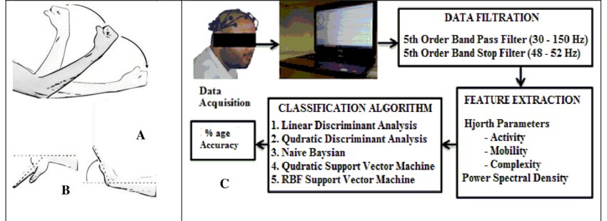
ResearchGate. (2026, March 3). Forearm extension and flexion EEG test protocol. https://www.researchgate.net/figure/A-Test-protocol-for-forearm-extension-and-flexion-for-recording-of_fig2_314188675
Science. (2026, March 3). Brain-computer interface research. https://www.science.org/doi/10.1126/science.abd0380
ScienceDirect. (2026, March 3). Brain-computer interface overview. https://www.sciencedirect.com/topics/neuroscience/brain-computer-interface
ScienceDirect. (2026, March 3). EEG and Alzheimer’s disease research. https://www.sciencedirect.com/science/article/pii/S2211383521003944
Springer. (2026, March 3). Brain-computer interface systems and applications. https://link.springer.com/chapter/10.1007/978-981-96-4512-1_2
Springer. (2026, March 3). EEG signal processing research. https://link.springer.com/article/10.1007/s12041-018-0962-4
World Health Organization. (2026, March 3). Dementia fact sheet. https://www.who.int/news-room/fact-sheets/detail/dementia
YouTube. (2026, March 3). Brain-computer interface explanation video. https://www.youtube.com/watch?v=8vKYAg9C8Jg
YouTube. (2026, March 3). EEG and BCI demonstration. https://www.youtube.com/watch?v=7hqwtT86DOs
YouTube. (2026, March 3). EEG brain activity explanation. https://www.youtube.com/watch?v=c_Fqt_WK-pk
YouTube. (2026, March 3). Neuron and brain signal explanation. https://www.youtube.com/watch?v=yJXTXN4xrI8
YouTube. (2026, March 3). Neuroscience and brain signals lecture.https://www.youtube.com/watch?v=v5gdH_Hydes
Goldberger, A. L., Amaral, L. A. N., Glass, L., Hausdorff, J. M., Ivanov, P. C., Mark, R. G., Mietus, J. E., Moody, G. B., Peng, C. K., & Stanley, H. E. (2026, March 3). EEG motor movement/imagery dataset. PhysioNet. https://www.physionet.org/content/eegmmidb/1.0.0/
Acknowledgement
I would like to thank my parents, friends, and superiors for their outmost support throughout my project. I would also Like to thank Mrs. O'Keefe for her feedback and suggestions and for the opportunity to participate in the fair.