Targeting MHC-1 Deficiency in Cancer With Peptide Immunotherapeutics Whilst Ensuring Red Blood Cell and MHC-1 Avoidance
Aiden Bell
Louis Riel School
Grade 9
Presentation
Hypothesis
The peptide LYQMELRYLAL with the S enantiomer will have the highest natural killer cell to red blood cell binding ratio, due to length of the peptide increasing its binding potential, and the S enantiomer being found more commonly in drugs targeting MHC-1 deficient cells.
Research
Key Formulas
Binding Free Energy (ΔG) from Dissociation Constant Kd
The relationship between binding free energy and affinity is: ΔG = RT ln Kd
Where:
-
ΔG = binding free energy (cal/mol or kcal/mol)
-
R = gas constant = 1.987 cal/(mol · K)
-
T = absolute temperature (Kelvin, usually 298 K ≈ 25°C)
-
Kd = dissociation constant (in M)
To convert to kcal/mol, divide by 1000. To convert kcal/kJ, multiply by 4.184 Example: Convert 1 µM Kd into ΔG
- Kd = 1 μM = 1 × 10⁻⁶ M
- R = 1.987 cal/(mol ⋅ K), T = 298 K
- RT = 1.987 × 298 ≈ 591.9 cal/mol
ΔG = (591.9) ln(1×10⁻⁶) ln(1×10⁻⁶) = −13.815 ΔG = 591.9 × (−13.815) ≈ −8175 cal/mol = −8.18 kcal/mol So a 1 µM binder has ΔG ≈ –8.2 kcal/mol.
Fold Difference Between Two Binding Energies (ΔG)
To compare two molecules: Kd2Kd1 = e ΔG2-ΔG1RT Where:
-
ΔG1 = binding energy of molecule 1
-
ΔG2 = binding energy of molecule 2
-
RT ≈ 591.9 cal/mol at 298 K
Example: Compare –6.82 vs –5.26 kcal/mol
- ΔG₁ = –6.82 kcal/mol = –6820 cal/mol
- ΔG₂ = –5.26 kcal/mol = –5260 cal/mol
ΔG2−ΔG1 = −5260 − (−6820) = 1560 cal/mol
Kd2Kd1 = e1560/591.9 ≈ e2.635 ≈ 13.9
Result: Molecule 1 (–6.82 kcal/mol) binds about 14× stronger than molecule 2 (–5.26 kcal/mol).
Amino Acids and Peptides
All 20 common amino acids are 𝛼-amino acids, having a carboxyl group and an amino group bonded to the 𝛼 carbon atom. (See figure below for the general structure of all but one naturally occurring amino acids). They differ mainly in their R groups, which can vary massively in  structure, size, charge, and can influence their solubility in water. These common amino acids have all been given three-letter abbreviations and one-letter symbols (see the table below).
 They are commonly used as shorthand (i.e., LYQLRY) to show the composition and structure of a peptide or protein. For all commonly found amino acids except for glycine (G), an 𝛼-carbon is bonded to a carboxyl group, an amino group, an R group, and a hydrogen atom (stereochemistry does still play a role in amino acids, as all amino acids have stereoisomers, i.e., alanine has L- and D-alanine for isomers). The 20 common amino acids are divided into 5 main classes based on their R groups, consisting of nonpolar, aliphatic R groups, aromatic R groups, polar uncharged R groups, positively charged R groups, and negatively charged R groups.
Nonpolar, aliphatic R groups are found within glycine, alanine, proline, valine, leucine, isoleucine, and methionine. They are nonpolar and hydrophobic, with the side chains of alanine, valine, leucine, and isoleucine clustering together within proteins, helping to stabilize the structures of proteins through the hydrophobicity of said amino acids. Glycine has the simplest structure, with its R group being a single hydrogen atom, and although it is grouped within the nonpolar amino acids, its small side chain makes very little contribution to interactions driven via the hydrophobic effect. Methionine is one of two sulphur-containing amino acids, and has a slightly nonpolar sulphide group in its side chain. Proline has an aliphatic side chain group with a cyclic structure. The secondary amino group of proline residues is held in a rigid arrangement, reducing the structural flexibility of any polypeptide groups containing proline.
Aromatic R groups are found within phenylalanine, tyrosine, and tryptophan, being relatively nonpolar and hydrophobic with their aromatic side chains. The hydroxyl group of tyrosine can form hydrogen bonds, an important functional group in some enzymes. Tyrosine and tryptophan are much more polar than phenylalanine due to the tyrosine hydroxyl group and the nitrogen of tryptophan’s indole ring. These amino acids with aromatic R groups absorb ultraviolet light, being used by researchers to characterize proteins.
Polar, uncharged R groups are found within serine, threonine, cysteine, asparagine, and glutamine. They are more hydrophilic than nonpolar R groups, containing functional groups that form hydrogen bonds with water molecules. Meanwhile, the polarity of each of these molecules is contributed by different groups. Serine and threonine’s hydroxyl group contributes to their polarity, while asparagine and glutamine’s amide groups contribute. Cysteine is quite different, being a weak acid with its polarity centred around the sulfhydryl group, allowing it to form weak bonds with oxygen and nitrogen.
Positively charged R groups are basic amino acids that include lysine, arginine, and histidine, and all have positive charges at pH 7.0. Lysine has a second primary amino group at the ɛ position on its aliphatic chain, arginine has a positively charged guanidinium group, and histidine has an aromatic imidazole group. Histidine is also the only natural amino acid that has an ionizable side chain with a pKa near neutrality, thus being able to have a polar charge or be uncharged at pH 7.0.
Negatively charged R groups consist of just two amino acids, aspartate and glutamate, each being slightly acidic. They both have a net negative charge at pH 7.0, and they each have a second carboxyl group.
They are commonly used as shorthand (i.e., LYQLRY) to show the composition and structure of a peptide or protein. For all commonly found amino acids except for glycine (G), an 𝛼-carbon is bonded to a carboxyl group, an amino group, an R group, and a hydrogen atom (stereochemistry does still play a role in amino acids, as all amino acids have stereoisomers, i.e., alanine has L- and D-alanine for isomers). The 20 common amino acids are divided into 5 main classes based on their R groups, consisting of nonpolar, aliphatic R groups, aromatic R groups, polar uncharged R groups, positively charged R groups, and negatively charged R groups.
Nonpolar, aliphatic R groups are found within glycine, alanine, proline, valine, leucine, isoleucine, and methionine. They are nonpolar and hydrophobic, with the side chains of alanine, valine, leucine, and isoleucine clustering together within proteins, helping to stabilize the structures of proteins through the hydrophobicity of said amino acids. Glycine has the simplest structure, with its R group being a single hydrogen atom, and although it is grouped within the nonpolar amino acids, its small side chain makes very little contribution to interactions driven via the hydrophobic effect. Methionine is one of two sulphur-containing amino acids, and has a slightly nonpolar sulphide group in its side chain. Proline has an aliphatic side chain group with a cyclic structure. The secondary amino group of proline residues is held in a rigid arrangement, reducing the structural flexibility of any polypeptide groups containing proline.
Aromatic R groups are found within phenylalanine, tyrosine, and tryptophan, being relatively nonpolar and hydrophobic with their aromatic side chains. The hydroxyl group of tyrosine can form hydrogen bonds, an important functional group in some enzymes. Tyrosine and tryptophan are much more polar than phenylalanine due to the tyrosine hydroxyl group and the nitrogen of tryptophan’s indole ring. These amino acids with aromatic R groups absorb ultraviolet light, being used by researchers to characterize proteins.
Polar, uncharged R groups are found within serine, threonine, cysteine, asparagine, and glutamine. They are more hydrophilic than nonpolar R groups, containing functional groups that form hydrogen bonds with water molecules. Meanwhile, the polarity of each of these molecules is contributed by different groups. Serine and threonine’s hydroxyl group contributes to their polarity, while asparagine and glutamine’s amide groups contribute. Cysteine is quite different, being a weak acid with its polarity centred around the sulfhydryl group, allowing it to form weak bonds with oxygen and nitrogen.
Positively charged R groups are basic amino acids that include lysine, arginine, and histidine, and all have positive charges at pH 7.0. Lysine has a second primary amino group at the ɛ position on its aliphatic chain, arginine has a positively charged guanidinium group, and histidine has an aromatic imidazole group. Histidine is also the only natural amino acid that has an ionizable side chain with a pKa near neutrality, thus being able to have a polar charge or be uncharged at pH 7.0.
Negatively charged R groups consist of just two amino acids, aspartate and glutamate, each being slightly acidic. They both have a net negative charge at pH 7.0, and they each have a second carboxyl group.
The Immune System
The immune system is a highly complex and coordinated network of cells, tissues, and signalling molecules that protects the body from infection, abnormal cells, and internal damage. Its primary roles include identifying pathogens, eliminating infected or transformed cells, and maintaining tolerance to healthy self-tissue. To accomplish this, the immune system must strike a delicate balance: it must respond aggressively to threats while avoiding excessive or misdirected activity that could harm the organism. This balance is achieved through multiple layers of regulation and communication between immune components. The immune system is broadly divided into two interconnected branches: the innate immune system and the adaptive immune system. The innate immune system provides rapid, non-specific defence and is the first line of protection against danger. It includes physical barriers such as the skin and mucosal surfaces, as well as immune cells like macrophages, neutrophils, dendritic cells, and natural killer (NK) cells. These components recognize conserved molecular patterns associated with pathogens or cellular stress rather than specific antigens. Because innate responses do not rely on prior exposure, they act within minutes to hours of detecting a threat. In contrast, the adaptive immune system is highly specific and capable of immunological memory. It relies primarily on lymphocytes — B cells and T cells — which recognize distinct molecular structures known as antigens. Adaptive immune responses take longer to develop but provide long-lasting protection. B cells produce antibodies that bind to extracellular targets, while T cells recognize antigenic peptides presented on major histocompatibility complex (MHC) molecules. Once activated, adaptive immune cells undergo clonal expansion, producing large numbers of cells tailored to a specific threat. Some of these cells persist as memory cells, enabling faster and stronger responses upon re-exposure. Communication between innate and adaptive immunity is essential for effective immune defence. Innate immune cells not only eliminate threats directly but also guide adaptive responses by presenting antigens and releasing signalling molecules called cytokines. Dendritic cells are particularly important in this role, as they capture antigens in peripheral tissues and migrate to lymph nodes to activate naïve T cells. This coordination ensures that immune responses are both immediate and precisely targeted. A critical function of the immune system is immune surveillance — the continuous monitoring of cells to detect abnormalities such as viral infection or malignant transformation. Healthy cells constantly display molecular indicators of their internal state on their surface. When these indicators change due to stress, mutation, or infection, immune cells can recognize the abnormal patterns and initiate elimination. This surveillance is especially important in preventing cancer, as cells frequently acquire mutations that could lead to uncontrolled growth if left unchecked. However, immune recognition is not based solely on detecting foreign molecules. The immune system must also distinguish between the healthy self and the dangerous non-self or altered-self. This discrimination is enforced through central and peripheral tolerance mechanisms. During immune cell development, cells that react strongly to self-antigens are eliminated or inactivated. Additional regulatory pathways suppress immune responses in healthy tissues. Failures in tolerance can lead to autoimmune diseases, while excessive tolerance can allow infections or tumours to persist. Inflammation is another key feature of immune activity. It is a coordinated response involving increased blood flow, immune cell recruitment, and release of signalling molecules at the site of injury or infection. While inflammation is essential for defence and tissue repair, chronic or uncontrolled inflammation can damage tissues and contribute to diseases such as cancer, cardiovascular disease, and neurodegeneration. The immune system, therefore, relies on precise regulatory mechanisms to initiate and resolve inflammatory responses appropriately. Cancer presents a unique challenge to the immune system because cancer cells originate from normal self-cells. As a result, tumours often resemble healthy tissue closely enough to evade immune detection. Additionally, cancer cells can actively suppress immune responses by altering signalling pathways, reducing antigen presentation, or creating immunosuppressive microenvironments. Understanding how the immune system interacts with cancer has become a major focus of modern biomedical research and has led to the development of immunotherapies that aim to restore or enhance immune function. Overall, the immune system is not a single entity but an integrated and adaptive network that continuously responds to internal and external challenges. Its ability to recognize subtle molecular changes, communicate across cell types, and regulate its own activity makes it one of the most sophisticated systems in biology. A detailed understanding of immune system function is essential for advancing treatments for infectious diseases, cancer, and immune-related disorders, as well as for the development of targeted therapies such as those involving natural killer cells.
MHC-I and the Immune System
Major Histocompatibility Complex class I (MHC-I) molecules are essential components of immune surveillance and play a central role in the immune system’s ability to detect infected or abnormal cells. These molecules are expressed on the surface of nearly all nucleated cells in the human body and function as molecular “display platforms” that present fragments of intracellular proteins to immune cells. By continuously sampling and displaying peptides derived from proteins synthesized within the cell, MHC-I molecules provide the immune system with real-time information about the cell’s internal state. The peptides presented by MHC-I are typically 8–10 amino acids in length and originate from proteins that are degraded by the proteasome, a cellular complex responsible for protein turnover. Once generated, these peptide fragments are transported into the endoplasmic reticulum by a specialized transporter known as TAP (Transporter Associated with Antigen Processing). Within the endoplasmic reticulum, peptides bind to newly synthesized MHC-I molecules, stabilizing them and allowing proper folding. The peptide–MHC-I complex is then transported to the cell surface, where it can be surveyed by immune cells. The primary immune cells responsible for monitoring MHC-I molecules are cytotoxic CD8⁺ T lymphocytes, which are part of the adaptive immune system. These T cells express T-cell receptors (TCRs) that recognize specific peptide–MHC-I combinations. Under normal conditions, peptides derived from self-proteins are presented, and T cells are tolerant to these signals. However, when a cell becomes infected by a virus or undergoes malignant transformation, abnormal peptides are generated and presented. Recognition of these non-self or altered-self peptides by CD8⁺ T cells triggers immune activation and targeted killing of the abnormal cell. This mechanism allows the immune system to detect threats that are hidden within cells, such as viruses that replicate intracellularly or cancer cells that harbour mutated proteins. MHC-I–mediated antigen presentation is therefore essential for adaptive immunity and long-term immune protection. Without functional MHC-I expression, CD8⁺ T cells are unable to recognize and eliminate infected or transformed cells, leading to increased susceptibility to disease. Despite its importance, MHC-I expression is frequently altered in disease states, particularly in cancer. Many tumour cells downregulate or completely lose MHC-I expression as a strategy to evade cytotoxic T-cell recognition. This immune evasion mechanism allows cancer cells to avoid adaptive immune responses even when they express abnormal proteins. Genetic mutations, epigenetic changes, or disruptions in antigen processing pathways can all contribute to reduced MHC-I expression in tumours. However, loss of MHC-I expression does not render cancer cells completely invisible to the immune system. Instead, it exposes them to surveillance by innate immune cells, particularly natural killer (NK) cells. NK cells rely on a balance of activating and inhibitory signals to determine whether a cell should be eliminated. One of the strongest inhibitory signals for NK cells comes from normal MHC-I expression. When MHC-I is absent or reduced, this inhibitory signal is lost, tipping the balance toward NK cell activation. This concept is known as the “missing-self” hypothesis and represents a crucial backup mechanism in immune defence. The interplay between MHC-I, T cells, and NK cells highlights the immune system’s redundancy and adaptability. While MHC-I enables highly specific adaptive immune responses, its loss activates innate immune pathways designed to compensate for immune evasion. This dual-layer surveillance system reduces the likelihood that abnormal cells can escape detection entirely. MHC-I molecules also play an important role in immune regulation beyond direct killing. The density and diversity of peptides presented influence immune tolerance, inflammation, and immune exhaustion. Chronic disease states, such as persistent viral infections or cancer, can alter MHC-I presentation patterns in ways that dampen immune responsiveness over time. Understanding these dynamics is essential for designing effective immunotherapies. In modern cancer research, restoring or manipulating MHC-I expression has become a key therapeutic strategy. Some treatments aim to increase MHC-I presentation to enhance T-cell recognition, while others deliberately exploit MHC-I deficiency to activate NK cell–mediated killing. The choice of strategy depends on tumour type, immune context, and treatment goals. As a result, MHC-I sits at the center of immune decision-making and represents a critical molecular interface between healthy self, diseased self, and immune response.
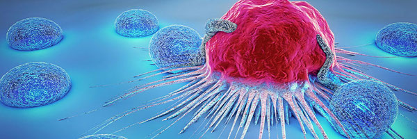
Natural Killer Cells
Natural killer (NK) cells are a specialized subset of lymphocytes that play a critical role in the innate immune system. Unlike adaptive immune cells such as T and B lymphocytes, NK cells do not require prior exposure to a specific antigen to become activated. Instead, they are capable of rapidly identifying and eliminating abnormal cells based on general indicators of cellular stress or transformation. This ability allows NK cells to act as an early defence mechanism against viral infections and cancer, often responding within hours of threat detection. NK cells originate in the bone marrow and circulate throughout the bloodstream and peripheral tissues. They are equipped with a diverse array of surface receptors that allow them to assess the health of surrounding cells. Rather than relying on a single activating signal, NK cells integrate signals from both activating and inhibitory receptors to determine whether a target cell should be destroyed. This signal integration model ensures that NK cells respond selectively to abnormal cells while sparing healthy tissue. One of the most important inhibitory signals for NK cells is the presence of normal MHC-I molecules on the surface of cells. Healthy cells express MHC-I, which binds to inhibitory receptors on NK cells and prevents activation. This interaction serves as a “do not kill” signal. When MHC-I expression is reduced or absent — a common feature of virus-infected cells and many cancer cells — this inhibitory signal is lost. The absence of MHC-I shifts the balance toward NK cell activation, a concept known as the missing-self hypothesis. This mechanism allows NK cells to target cells that evade cytotoxic T lymphocytes by downregulating antigen presentation. In addition to sensing missing MHC-I, NK cells recognize activating signals associated with cellular stress. These signals include stress-induced ligands that are upregulated in response to DNA damage, infection, or malignant transformation. When activating signals outweigh inhibitory ones, NK cells initiate cytotoxic responses. This dual-signal requirement ensures specificity and prevents unintended destruction of healthy cells. Once activated, NK cells eliminate target cells through multiple mechanisms. One primary method involves the release of cytotoxic granules containing perforin and granzymes. Perforin forms pores in the target cell membrane, allowing granzymes to enter and trigger programmed cell death (apoptosis). NK cells can also induce apoptosis through receptor-mediated pathways by expressing death-inducing ligands that bind to receptors on the target cell surface. These mechanisms enable NK cells to kill efficiently while minimizing inflammation and collateral tissue damage. Beyond direct cytotoxicity, NK cells play an important regulatory role in shaping immune responses. They secrete cytokines such as interferon-gamma (IFN-γ), which enhances antigen presentation, activates macrophages, and supports adaptive immune responses. Through cytokine signalling, NK cells help coordinate interactions between innate and adaptive immunity, influencing the behaviour of T cells, dendritic cells, and other immune populations. This regulatory role highlights that NK cells are not only killers but also key immune communicators. NK cells are particularly important in the context of cancer because tumour cells frequently develop mechanisms to escape adaptive immune detection. Many cancers downregulate MHC-I expression to avoid recognition by cytotoxic T cells, inadvertently making themselves more susceptible to NK cell–mediated killing. However, tumours can also evolve strategies to suppress NK cell activity, such as secreting inhibitory cytokines or altering the expression of activating ligands. These evasion tactics contribute to tumour progression and immune resistance. The functional state of NK cells can vary significantly depending on their environment. Factors such as cytokine availability, metabolic conditions, and interactions with other immune cells influence NK cell responsiveness. In chronic disease states, including cancer, NK cells may become functionally impaired or “exhausted,” reducing their cytotoxic capacity. Understanding these regulatory influences is critical for designing therapies that aim to restore or enhance NK cell function. Due to their rapid response, MHC-I sensitivity, and ability to target transformed cells, NK cells occupy a unique and essential position in immune surveillance. They serve as a bridge between innate and adaptive immunity and act as a safeguard against immune evasion strategies employed by pathogens and tumours. Their biology makes them particularly attractive targets for immunotherapeutic intervention, especially in cancers characterized by altered antigen presentation.
Natural Killer Cell–Focused Therapies
Natural killer (NK) cell–focused therapies are an emerging class of immunotherapies that aim to harness or enhance the innate immune system’s ability to recognize and eliminate cancer cells. Unlike adaptive immune therapies that depend on antigen specificity and prior immune priming, NK cell–based approaches take advantage of NK cells’ natural capacity to detect cellular stress and loss of normal self-markers such as MHC-I. This makes NK cell therapies particularly promising for tumours that evade T cell–mediated immunity through antigen loss or reduced antigen presentation. One of the earliest NK cell–based strategies involves the isolation and expansion of NK cells outside the body, followed by reinfusion into patients. These adoptive NK cell therapies can use either autologous NK cells, which are derived from the patient, or allogeneic NK cells obtained from a donor. Allogeneic NK cells are often favoured because they may be more active against tumour cells due to reduced inhibitory signalling. Once infused, expanded NK cells can circulate and target cancer cells directly through cytotoxic mechanisms. Advances in genetic engineering have further expanded the potential of NK cell therapies. NK cells can be modified to express engineered receptors that improve their ability to recognize tumour-specific markers. These engineered NK cells share conceptual similarities with CAR-T cells but differ in important ways. NK cells are less likely to cause severe immune overactivation and inflammatory side effects, making them potentially safer for clinical use. Additionally, engineered NK cells can retain their innate ability to detect MHC-I–deficient cells, providing a dual mechanism of tumour recognition. Another major area of NK cell–focused therapy involves modulating the signalling pathways that regulate NK cell activity. Tumour cells often exploit inhibitory receptors on NK cells to suppress their cytotoxic function. Therapeutic agents that block these inhibitory interactions can effectively “release the brakes” on NK cells, allowing them to respond more aggressively to cancer cells. Conversely, some therapies aim to enhance activating signals by increasing the expression of stress ligands on tumour cells or stimulating NK cells with cytokines. Cytokine-based therapies are also being explored to support NK cell survival, proliferation, and activation in vivo. Certain cytokines promote NK cell expansion and enhance their cytotoxic capacity, improving anti-tumour responses. However, systemic cytokine administration must be carefully controlled, as excessive immune stimulation can lead to toxicity. Current research focuses on localized or targeted cytokine delivery to maximize therapeutic benefit while minimizing side effects. NK cell therapies are especially relevant for cancers that exhibit low or absent MHC-I expression. Because such tumours often resist conventional T cell–based immunotherapies, NK cells provide an alternative immune pathway capable of targeting these evasive cancer cells. This makes NK-focused strategies valuable both as standalone treatments and in combination with other immunotherapies. Combination approaches may enhance overall immune effectiveness by engaging multiple immune mechanisms simultaneously. Despite their promise, NK cell–focused therapies face several challenges. NK cells can have limited persistence in the body after infusion, and the tumour microenvironment may suppress NK cell function through inhibitory signals and metabolic constraints. Researchers are actively investigating ways to improve NK cell longevity, trafficking, and resistance to immunosuppressive conditions. These efforts include genetic modifications, metabolic optimization, and combination treatment strategies. Overall, NK cell–focused therapies represent a rapidly advancing frontier in cancer immunotherapy. By leveraging innate immune mechanisms and targeting vulnerabilities in immune-evasive tumours, these approaches offer a complementary alternative to existing treatments. Continued research into NK cell biology, regulation, and therapeutic engineering is expected to expand their clinical impact and improve outcomes for patients with resistant or advanced cancers.
Molecular Docking and Binding Energetics
Molecular docking is a computational technique used to predict how two molecules interact in three-dimensional space, most commonly a ligand and a target protein. In biomedical research, docking is widely used to model how drug candidates bind to receptors, enzymes, or immune-related proteins. By simulating these interactions in silico, researchers can evaluate potential therapeutic molecules efficiently before committing to laboratory experiments. Molecular docking is especially valuable in early-stage drug discovery, where it allows thousands of candidate compounds to be screened rapidly and cost-effectively. At its core, molecular docking attempts to answer two fundamental questions: how a molecule binds to a target, and how strongly it binds. Docking algorithms generate multiple possible binding orientations, known as poses, by positioning the ligand within the protein’s binding site and allowing limited conformational flexibility. Each pose is evaluated using a scoring function that estimates the favorability of the interaction. These scoring functions are designed to approximate the physical forces that govern molecular binding. Binding energetics refers to the change in free energy that occurs when a ligand binds to a protein. This binding free energy reflects the balance between stabilizing and destabilizing interactions. Favourable contributions include hydrogen bonds, electrostatic interactions, hydrophobic effects, and van der Waals forces, while unfavourable contributions may arise from steric clashes or loss of conformational freedom. In docking studies, more negative binding energy values generally indicate stronger and more stable interactions, suggesting a higher likelihood that binding will occur under physiological conditions. Hydrogen bonding plays a critical role in molecular recognition, particularly in biological systems where specificity is essential. These interactions occur when hydrogen atoms are shared between electronegative atoms such as oxygen or nitrogen, stabilizing ligand–protein complexes. Electrostatic interactions further contribute to binding by attracting oppositely charged regions of molecules. Hydrophobic interactions, though not based on direct attraction, are driven by the tendency of nonpolar groups to avoid water, effectively stabilizing the ligand within hydrophobic pockets of the protein. Molecular docking is particularly useful for studying peptide–protein interactions, which are often difficult to characterize experimentally due to their size and flexibility. Peptides can adopt multiple conformations, and their binding modes are influenced by stereochemistry, backbone flexibility, and side-chain orientation. Computational docking allows researchers to explore how different peptide sequences or stereochemical configurations interact with immune receptors or signalling proteins, providing insight into which designs are most promising. Despite its usefulness, molecular docking relies on approximations that limit its predictive accuracy. Most docking algorithms treat the protein as relatively rigid, even though proteins are dynamic molecules that undergo conformational changes during binding. Solvent effects, temperature, and long-range interactions are often simplified or partially ignored. As a result, docking scores should be interpreted as relative comparisons rather than exact measurements of binding strength. Another limitation lies in the scoring functions themselves. Different docking programs may produce different binding energy values for the same interaction because they weigh energetic contributions differently. Additionally, binding affinity alone does not guarantee biological effectiveness. A molecule may bind strongly to a target protein but fail to function in vivo due to instability, poor bioavailability, or unintended interactions. For this reason, molecular docking is best viewed as a hypothesis-generating tool rather than definitive proof of therapeutic potential. In the context of immunological research, docking is often used to study interactions involving immune receptors, antigen-presenting molecules, and immune-modulating ligands. For example, docking can help predict how peptides bind to immune-related proteins or how therapeutic molecules interact with receptors involved in immune activation or inhibition. These predictions can guide rational design by identifying key residues involved in binding and suggesting modifications that improve specificity or affinity. Molecular docking also plays an important role in reducing experimental burden. By narrowing down candidate molecules computationally, researchers can focus laboratory resources on the most promising designs. This is especially important in complex systems such as immune signalling, where experimental validation can be time-consuming and costly. When combined with experimental data, docking results provide a powerful framework for understanding molecular interactions and optimizing therapeutic candidates. Overall, molecular docking and binding energy analysis form a critical bridge between theoretical chemistry and practical biomedical application. While computational predictions must be interpreted with caution, they offer valuable insights into molecular behaviour that would otherwise be difficult to obtain. In modern drug discovery and immunotherapy research, docking serves as an essential tool for guiding rational design and accelerating scientific progress.
Stereochemistry in Medicine
Stereochemistry is the study of how the three-dimensional arrangement of atoms within a molecule influences its chemical and biological behaviour. In biological systems, stereochemistry is particularly important because most biomolecules, including proteins, enzymes, and receptors, are inherently three-dimensional and chiral. Chirality refers to the property of a molecule that makes it non-superimposable on its mirror image, similar to the relationship between left and right hands. These mirror-image forms are known as enantiomers. Although enantiomers share the same molecular formula and connectivity, their different spatial arrangements can lead to dramatically different interactions with biological targets. In medicine, stereochemistry often determines whether a drug is effective, inactive, or harmful. Because biological receptors are chiral, they can distinguish between different stereoisomers of the same compound. One stereoisomer may fit precisely into a binding site and trigger a desired biological response, while another may bind weakly or not at all. In some cases, an incorrect stereochemical configuration can cause unintended interactions with other proteins, leading to side effects or toxicity. This makes stereochemical control a critical consideration in drug development. Historically, many drugs were administered as racemic mixtures containing equal amounts of two enantiomers. Over time, it became clear that this approach could reduce efficacy or increase risk. Advances in chemical synthesis and separation techniques have since allowed researchers to produce single-enantiomer drugs with greater precision. Modern pharmaceuticals increasingly rely on stereochemically pure compounds to improve the safety, potency, and predictability of biological effects. Stereochemistry is particularly important in peptide-based therapeutics. Amino acids, the building blocks of peptides and proteins, are chiral molecules, and naturally occurring proteins are composed almost exclusively of one stereochemical form. The spatial orientation of amino acid side chains affects peptide folding, stability, and binding interactions. Even small stereochemical changes within a peptide sequence can alter its conformation and disrupt its ability to interact with a target protein. As a result, stereochemical considerations are essential when designing peptides intended to bind immune receptors or signalling molecules. In computational drug design, stereochemistry has a direct influence on molecular docking results and binding energy predictions. Docking algorithms evaluate how well a molecule’s three-dimensional shape complements a protein’s binding site. Two stereoisomers of the same compound can produce significantly different docking poses and predicted binding affinities due to differences in orientation and functional group positioning. Accurate modelling of stereochemistry is therefore essential for generating meaningful computational predictions. Stereochemistry also affects pharmacokinetic properties such as metabolism and distribution. Enzymes responsible for drug metabolism are stereoselective and may process one stereoisomer more rapidly than another. This can influence how long a drug remains active in the body and how it is eliminated. Understanding these effects allows researchers to design molecules with improved stability and predictable behaviour in vivo. Overall, stereochemistry is a foundational concept in modern medicine and drug discovery. It links molecular structure to biological function and plays a key role in determining drug efficacy, safety, and specificity. In computational and experimental research alike, careful attention to stereochemical detail is essential for designing therapeutic molecules that interact reliably and selectively with biological targets.
Peptide Therapeutics
Peptide therapeutics are a class of drugs composed of short chains of amino acids designed to influence biological processes with high specificity. Because peptides are constructed from the same building blocks as proteins, they can closely mimic natural signalling molecules such as hormones, cytokines, and receptor ligands. This structural similarity allows peptide therapeutics to interact selectively with biological targets, making them particularly useful in treating diseases that involve precise molecular signalling, including cancer, metabolic disorders, and immune-related conditions. One of the major advantages of peptide therapeutics is their target specificity. Peptides can be engineered to recognize unique structural features on proteins that are difficult for traditional small-molecule drugs to target. This specificity often reduces off-target effects and improves safety. In immunology, peptides are especially valuable because immune signalling relies heavily on protein–protein interactions. Therapeutic peptides can be designed to activate, inhibit, or modulate immune pathways with a high degree of precision. Despite these advantages, peptide therapeutics face significant challenges related to stability and delivery. In the bloodstream, peptides are susceptible to degradation by proteolytic enzymes, which can rapidly reduce their effectiveness. Additionally, peptides often have limited ability to cross cell membranes and may be cleared quickly from circulation. These limitations have historically restricted the use of peptide drugs, particularly for systemic applications. To overcome these challenges, researchers employ a variety of peptide optimization strategies. Structural modifications such as cyclization, backbone stabilization, and incorporation of non-natural amino acids can significantly improve resistance to enzymatic degradation. Stereochemical optimization also plays a role, as altering the spatial configuration of amino acids can enhance stability without compromising biological activity. These modifications allow peptide therapeutics to maintain their functional properties while achieving improved pharmacokinetic performance. Computational tools play an increasingly important role in peptide therapeutic development. Molecular modelling and docking simulations allow researchers to predict how peptide sequences will fold and interact with target proteins. By evaluating binding affinity, specificity, and structural compatibility in silico, researchers can refine peptide designs before synthesis and experimental testing. This approach reduces development time and focuses experimental efforts on the most promising candidates. Peptide therapeutics are particularly relevant in immune-based treatments due to their ability to interact with immune receptors and signalling molecules. They can be designed to selectively target immune cells or modulate immune responses in disease-specific contexts. For example, peptides may be engineered to preferentially interact with receptors expressed on immune cells involved in tumour surveillance. This selectivity makes peptides a powerful platform for immunotherapy research. Overall, peptide therapeutics represent a rapidly growing field that bridges molecular biology, chemistry, and medicine. Advances in peptide design, stabilization, and computational modelling have expanded their clinical potential and broadened their range of applications. As drug discovery increasingly moves toward precision medicine, peptide therapeutics offer a versatile and effective approach for targeting complex biological systems.
Drug-Likeness and ADMET
In drug development, strong target binding alone is not sufficient for a compound to become a successful therapeutic. A molecule must also possess properties that allow it to function effectively and safely within the human body. The concept of drug-likeness refers to a set of chemical and physical characteristics that influence whether a compound is suitable for use as a drug. These characteristics include molecular size, solubility, polarity, stability, and chemical reactivity. Compounds that fall outside acceptable ranges for these properties may fail despite promising biological activity. ADMET — an acronym for Absorption, Distribution, Metabolism, Excretion, and Toxicity — describes how a compound behaves after it enters the body. Absorption refers to how efficiently a drug enters the bloodstream, while distribution describes how it spreads through tissues and organs. Metabolism involves chemical modification of the drug by enzymes, primarily in the liver, and excretion refers to how the drug is removed from the body. Toxicity encompasses any harmful effects the compound may have on healthy tissues. Together, these factors determine whether a drug reaches its target at therapeutic levels without causing unacceptable side effects. Peptide therapeutics present unique challenges in terms of drug-likeness and ADMET. Compared to small-molecule drugs, peptides are often larger, more flexible, and more susceptible to enzymatic degradation. These properties can reduce oral bioavailability and shorten circulation time in the bloodstream. As a result, many peptide drugs require alternative delivery methods or structural optimization to achieve effective concentrations in vivo. Evaluating ADMET properties early in development is therefore especially important for peptide-based candidates. Computational ADMET prediction tools are widely used to assess drug-likeness before experimental testing. These tools analyze molecular features such as charge distribution, hydrophobicity, and molecular weight to estimate how a compound will behave in biological systems. While these predictions are approximations, they allow researchers to identify potential issues early and refine molecular designs accordingly. In computational drug discovery projects, ADMET screening helps prioritize candidates that balance biological potency with practical feasibility. Metabolism is a particularly important consideration in peptide drug design. Enzymes in the blood and tissues can rapidly degrade peptides into inactive fragments. Structural modifications, such as incorporating non-natural amino acids or stabilizing the peptide backbone, can slow metabolic breakdown and extend therapeutic activity. These modifications must be carefully designed to preserve target binding while improving metabolic stability. Toxicity assessment is also essential, especially for immune-targeted therapies. A molecule that overstimulates the immune system or interacts with unintended targets can cause serious adverse effects. Computational toxicity predictions help identify molecular features associated with immune overactivation, off-target binding, or cellular damage. Although these tools cannot fully replace experimental safety testing, they provide valuable guidance during early design stages. Overall, drug-likeness and ADMET considerations form a critical bridge between theoretical molecular design and real-world medical application. By evaluating how a compound behaves within the body, researchers can ensure that promising molecular interactions translate into viable therapies. In peptide-based and immune-focused drug discovery, balancing binding strength with favourable ADMET properties is essential for successful therapeutic development.
Protein Structure Basics
Proteins are essential biological macromolecules that carry out most cellular functions, including catalysis, signalling, structural support, and immune recognition. They are composed of amino acids linked together by peptide bonds to form polypeptide chains. The specific order of amino acids, known as the primary structure, is determined by genetic information and ultimately dictates how the protein folds and functions. Even a single change in amino acid sequence can alter protein behaviour, stability, or interaction with other molecules. Protein structure is organized into four hierarchical levels. The primary structure is the linear amino acid sequence. The secondary structure refers to local folding patterns such as alpha helices and beta sheets, which arise from hydrogen bonding between backbone atoms. These elements form predictable structural motifs that contribute to overall stability. The tertiary structure describes the full three-dimensional shape of a single polypeptide chain, resulting from interactions between amino acid side chains, including hydrogen bonds, ionic interactions, hydrophobic effects, and disulphide bonds. Some proteins also exhibit quaternary structure, in which multiple polypeptide chains assemble into a functional complex. The three-dimensional structure of a protein determines how it interacts with other molecules. Binding sites, active sites, and interaction surfaces are shaped by the protein’s folding pattern and chemical properties. In immune biology, protein structure is especially important because immune recognition depends on precise molecular interactions. Receptors on immune cells recognize specific structural features of other proteins or peptide fragments, and even subtle conformational differences can determine whether binding occurs. Protein flexibility is another important aspect of structure. While proteins are often depicted as rigid shapes, they are dynamic molecules that undergo constant motion. Conformational changes can occur upon ligand binding, during signalling, or in response to environmental conditions such as pH and temperature. This flexibility allows proteins to perform complex functions but also complicates efforts to model them computationally. Many computational approaches approximate proteins as relatively rigid, which can affect prediction accuracy. In drug discovery and peptide design, understanding protein structure is critical for identifying suitable binding targets. Structural data obtained through techniques such as X-ray crystallography, nuclear magnetic resonance (NMR) spectroscopy, and cryo-electron microscopy provide detailed models of protein architecture. These structures serve as the foundation for molecular docking studies, where candidate molecules are tested for compatibility with binding sites. Accurate structural information increases the likelihood that computational predictions reflect real biological interactions. Protein structure is also closely linked to disease. Misfolded proteins can lose function or form harmful aggregates, contributing to conditions such as neurodegenerative disorders and cancer. In cancer biology, mutations may alter protein structure in ways that promote uncontrolled growth or immune evasion. Understanding these structural changes helps researchers design therapeutic molecules that specifically target diseased cells while sparing healthy tissue. Overall, protein structure provides the physical basis for molecular function in biology. From immune recognition to drug binding, the shape and flexibility of proteins govern how biological systems operate. A solid understanding of protein structure is therefore essential for interpreting molecular docking results, designing peptide therapeutics, and developing immune-focused treatments.
Computational Limits
Computational modelling plays a central role in modern drug discovery by allowing researchers to explore molecular interactions efficiently and at low cost. Techniques such as molecular docking, binding energy estimation, and ADMET prediction enable the screening of large numbers of compounds before experimental testing. However, despite their usefulness, computational approaches have inherent limitations that must be acknowledged when interpreting results. Understanding these limits is essential for evaluating the reliability and scope of in silico findings. One major limitation of computational modelling is the reliance on simplifications and assumptions. Many docking algorithms treat proteins as rigid or only partially flexible structures, even though proteins are dynamic molecules that constantly change conformation in biological environments. This approximation can lead to inaccurate predictions of binding orientation or affinity, particularly for flexible targets such as immune receptors and peptide–protein interfaces. As a result, strong predicted binding in silico does not always translate into effective binding in vivo. Another constraint is the accuracy of scoring functions used to estimate binding strength. Docking programs approximate complex physical interactions using mathematical models that balance speed and precision. These scoring functions may not fully capture important contributions such as solvent effects, long-range electrostatics, or entropic changes upon binding. For peptide-based interactions, which often involve multiple contact points and conformational adjustments, these limitations can significantly affect predicted binding energies. Computational power also restricts the level of detail that can be realistically modelled. High-accuracy simulations, such as molecular dynamics with explicit solvent and full protein flexibility, require substantial computational resources and time. As a result, many student-level and early-stage research projects rely on faster but less detailed methods. While these approaches are valuable for hypothesis generation and comparative analysis, they cannot replace experimental validation. Biological complexity further limits computational predictability. Living systems involve numerous interacting variables, including competing ligands, cellular membranes, enzymatic degradation, and immune regulation. Computational models typically examine isolated interactions under idealized conditions, which may not reflect the crowded and variable environment of the human body. This is particularly relevant in immune-focused therapies, where small changes in signalling balance can produce large biological effects. Data quality is another important factor. Computational models depend on existing structural and biochemical data, which may be incomplete or biased toward well-studied proteins. Errors in protein structures, missing regions, or inaccurate protonation states can propagate through docking and simulation workflows, affecting results. For novel or less-characterized immune targets, these limitations become more pronounced. Despite these constraints, computational methods remain powerful tools when used appropriately. They are most effective for narrowing candidate pools, identifying trends, and guiding experimental design rather than producing definitive answers. In science fair–level research, acknowledging computational limits demonstrates scientific rigour and an understanding of how theoretical modelling fits into the broader research pipeline. In summary, computational modelling provides valuable insights into molecular interactions but cannot fully replicate biological reality. Recognizing the limitations of in silico methods strengthens the interpretation of results and highlights the importance of experimental validation. By combining computational predictions with biological knowledge, researchers can use these tools responsibly and effectively in the development of immune-targeted therapeutics.
Sources
Dettmer, P. (2021). Immune: A journey into the mysterious system that keeps you alive. Random House. Nelson, D. L., Cox, M. M., Hoskins, A. A., & Lehninger, A. L. (2023). Lehninger Principles of Biochemistry (7th ed.). Janeway, C. (2011). Janeway’s Immunobiology (8th ed.). Fosgerau, K., & Hoffmann, T. (2015). Peptide therapeutics: Current status and future directions. Drug Discovery Today, 20(1), 122–128. https://www.sciencedirect.com/science/article/pii/S1359644614003997?via%3Dihub Lau, J. L., & Dunn, M. K. (2018). Therapeutic peptides: Historical perspectives, current development trends, and future directions. Bioorganic & Medicinal Chemistry, 26(10), 2700–2707. https://www.sciencedirect.com/science/article/pii/S0968089617310222?via%3Dihub Meng, X. Y., Zhang, H. X., Mezei, M., & Cui, M. (2011). Molecular docking: A powerful approach for structure-based drug discovery. Current Computer-Aided Drug Design, 7(2), 146–157. https://www.eurekaselect.com/article/19093 Kitchen, D. B., Decornez, H., Furr, J. R., & Bajorath, J. (2004). Docking and scoring in virtual screening for drug discovery: Methods and applications. Nature Reviews Drug Discovery, 3(11), 935–949. https://www.nature.com/articles/nrd1549 Lipinski, C. A., Lombardo, F., Dominy, B. W., & Feeney, P. J. (2001). Experimental and computational approaches to estimate solubility and permeability in drug discovery and development settings. Advanced Drug Delivery Reviews, 46(1–3), 3–26.https://www.sciencedirect.com/science/article/abs/pii/S0169409X00001290?via%3Dihub van de Waterbeemd, H., & Gifford, E. (2003). ADMET in silico modelling: Towards prediction paradise? Nature Reviews Drug Discovery, 2(3), 192–204. https://www.nature.com/articles/nrd1032 Warren, G. L., Andrews, C. W., Capelli, A. M., Clarke, B., LaLonde, J., Lambert, M. H., … Head, M. S. (2006). A critical assessment of docking programs and scoring functions. Journal of Medicinal Chemistry, 49(20), 5912–5931. https://pubs.acs.org/doi/10.1021/jm050362n Jorgensen, W. L. (2004). The many roles of computation in drug discovery. Science, 303(5665), 1813–1818. https://www.science.org/doi/10.1126/science.1096361
Variables
Manipulated: The construction of the peptides (i.e., LYQLRY, LYFLRY, etc.) Responding: The ability of the peptide to bind to 1T2Q (red blood cell) and 8TLZ (NK receptor) Controlled:
- Temperature (37 °C)
- Same solvent model (implicit water)
- Program used for peptide design (PyMol/ChimeraX)
- Program used for peptide binding (AutoDock Vina)
- Same program versions used across trials
- Same receptors used
- Same binding location (i.e., I can’t have one peptide bind at the top and the other at the bottom of the ligand)
- Same protein preparation procedure
- Only the target receptor, nothing else
- Same simulation number
- Same affinity output (ΔG vs Kd vs M)
- Same selection criteria
Procedure
Materials:
- Computer with Internet access
- ChimeraX (generate peptides/imaging)
- PyMol (R/S enantiomers)
- AutoDock Tools (prep peptides for Vina)
- AutoDock Vina (test docking)
- Protein Structures from PDB
- 3D printer with printing material to print peptide models
Procedure:
- Identify NK cell activating receptor (NKG2D) using PDB.
- Download receptor structure (PDB ID: 4S0U).
- Open the receptor in AutoDock Tools.
- Delete all water molecules.
- Add polar hydrogens.
- Compute Gasteiger charges.
- Go to ligand and select “choose molecule,” then choose 4S0U.
- Save receptor as a PDBQT file for AutoDock Vina.
- Open ChimeraX.
- Open tools → structure editing → build structure → peptide.
- Enter the sequence (eg, LYQLRY).
- Click build.
- Save peptide.
- In Pymol, make R enantiomer (use this command: transform_selection all, (-1,0,0, 0,1,0, 0,0,1, 0,0,0)).
- Save molecule.
- Repeat steps 9 - 15 for all peptides (I made 6).
- Download a red blood cell membrane protein (glycophorin A) from PDB (PDB ID:1T2Q).
- Prepare in AutoDock Tools, same as receptor (delete water, add polar hydrogens, compute Gasteiger charges, choose molecule).
- Save as a PDBQT to test off-target binding.
- Open AutoDock Vina.
- Load NK receptor PDBQT as receptor.
- Load peptide as ligand.
- Define the grid box around the receptor binding site.
- Run docking and record binding affinity (kcal/mol).
- Repeat procedure with the RBC protein as the receptor.
- Repeat steps 20 - 25 for all peptides and enantiomers.
- Compare binding energies for the NK receptor vs RBC proteins.
- Calculate the ratio of binding using this formula: Kd2Kd1 = e ΔG2-ΔG1RT (You want it to be ideally over 10:1, as most drugs have a similar ratio, if not higher)(you can convert to molar as well, if you want).
- Now check for drug likeness using Lipinski’s Rule of 5 (Molecular Weight < 500 Da, LogP < 5, ≤5 Hydrogen-bond donors, ≤10 Hydrogen-bond acceptors). This can be done using SwissADME or manually, though manually takes much longer.
- Record observations.
- If necessary, use AI to mutate the peptide and repeat steps 9-30 for a higher efficacy.
Observations


| Peptide | Binding Strength to 8TLZ
(NK) at 310 K (M) | Binding Strength to 1T2Q
(RBC) at 310 K (M) | Binding Strength to 6JTN (MHC-1) at 310 K (M) | Binding Strength to 6JTP (MHC-1) at 310 K (M) | Binding Strength to 6P64 (MHC-1) at 310 K (M) | Binding Strength to 6TRN (MHC-1) at 310 K (M) | Binding Strength to 6TRO (MHC-1) at 310 K (M) | Binding Strength to 6ULI (MHC-1) at 310 K (M) | Average Binding Strength To MHC | Ratio of Binding Strength to 8TLZ vs 1T2Q | Ratio of Binding Strength to 8TLZ vs MHC-1 (Mean) | | ------- | ----------------------------------------- | ------------------------------------------ | --------------------------------------------- | --------------------------------------------- | --------------------------------------------- | --------------------------------------------- | --------------------------------------------- | --------------------------------------------- | ------------------------------- | ----------------------------------------- | ------------------------------------------------- | | Cyclo(LYQFRYLAL) - L | 600 nM | 21 µM | 2.9 µM | 5.1 µM | 4.3 µM | 11 µM | 19 µM | 8 µM | 8.1 µM | 36:1 | 13:1 | | Cyclo(LYFWRYLAL) - L | 260 nM | 9.3 µM | 2.7 µM | 4.3 µM | 3.7 µM | 8 µM | 13 µM | 5.1 µM | 5.8 µM | 50:1 | 22:1 | | Cyclo(LYQFDWRYLPL) - L | 140 nM | 4.3 µM | 1.9 µM | 3.2 µM | 2.7 µM | 5.1 µM | 6.8 µM | 4.3 µM | 3.5 µM | 25:1 | 28:1 | | LYQLRY - D | 50.1 µM | 474 µM | N/A | N/A | N/A | N/A | N/A | N/A | N/A | \~9.5:1 | N/A | | LYQLRY - L | 9.43 mM | 27.4 mM | N/A | N/A | N/A | N/A | N/A | N/A | N/A | \~2.95:1 | N/A | | LYQLRYLAL - D | 22.3 µM | 267 µM | N/A | N/A | N/A | N/A | N/A | N/A | N/A | \~12:1 | N/A | | LYQLRYLAL - L | 3.85 mM | 13.9 mM | N/A | N/A | N/A | N/A | N/A | N/A | N/A | \~3.58:1 | N/A | | LYFLRY - D | 73.9 µM | 534 µM | N/A | N/A | N/A | N/A | N/A | N/A | N/A | \~7.2:1 | N/A | | LYFLRY - L | 8.52 mM | 23.1 mM | N/A | N/A | N/A | N/A | N/A | N/A | N/A | \~2.71:1 | N/A | | LYFLRYLAL - D | 38.9 µM | 295 µM | N/A | N/A | N/A | N/A | N/A | N/A | N/A | \~7.6:1 | N/A | | LYFLRYLAL - L | 9.43 mM | 23.9 mM | N/A | N/A | N/A | N/A | N/A | N/A | N/A | \~2.55:1 | N/A | | LYQMELRY - D | 28.7 µM | 311 µM | N/A | N/A | N/A | N/A | N/A | N/A | N/A | \~10.8:1 | N/A | | LYQMELRY - L | 5.98 mM | 13.9 mM | N/A | N/A | N/A | N/A | N/A | N/A | N/A | \~2.32:1 | N/A | | LYQMELRYLAL - D | 9.89 µM | 138 µM | N/A | N/A | N/A | N/A | N/A | N/A | N/A | \~14:1 | N/A | | LYQMELRYLAL - L | 3.54 mM | 12.8 mM | N/A | N/A | N/A | N/A | N/A | N/A | N/A | \~2.58:1 | N/A |
| Peptide | Length (Number of Amino Acids) | Molecular Weight (Daltons) | Net Charge (approx. at pH 7.4) | Hydrophobic Residues | Aromatic Residues | Predicted Isoelectric Point (pI) | GRAVY (Grand Average of Hydropathy) | Molecular Weight ≤500 Da | Hydrogen Bond Donors ≤5 | Hydrogen Bond Acceptors ≤10 | LogP ≤5 | Violations |
|---|---|---|---|---|---|---|---|---|---|---|---|---|
| LYQLRY | 6 | 854.97 | 0.8 | 4 | 2 | \~9.1 | 0.567 | No | Yes | Yes | Yes | 1 (Molecular Weight) |
| LYQLRYLAL | 9 | 1152.34 | 0.8 | 7 | 2 | \~9.1 | 0.611 | No | No | No | Yes | 3 (Molecular Weight, Hydrogen Bond Donors, Hydrogen Bond Acceptors) |
| LYFLRY | 6 | 874.01 | 0.8 | 5 | 3 | \~9.1 | 0.7 | No | Yes | Yes | Yes | 1 (Molecular Weight) |
| LYFLRYLAL | 9 | 1171.38 | 0.8 | 8 | 3 | \~9.1 | 0.689 | No | No | No | Yes | 3 (Molecular Weight, Hydrogen Bond Donors, Hydrogen Bond Acceptors) |
| LYQMELRY | 7 | 1005.2 | 0.8 | 6 | 3 | \~9.0 | 0.671 | No | No | No | Yes | 2 (Molecular Weight, Hydrogen Bond Acceptors borderline) |
| LYQMELRYLAL | 10 | 1302.57 | 0.8 | 9 | 3 | \~9.0 | 0.67 | No | No | No | Yes | 3 (Molecular Weight, Hydrogen Bond Donors, Hydrogen Bond Acceptors) |
| Cyclo(LYQFRYLAL) | 9 | 1149.63 | 1 | 7 | 3 | \~10.1 | 0.6 | No | No | No | Yes | 3 (Molecular Weight, Hydrogen Bond Donors, Hydrogen Bond Acceptors) |
| Cyclo(LYFWRYLAL) | 9 | 1197.65 | 1 | 8 | 4 | \~10.1 | 0.89 | No | No | No | Yes | 3 (Molecular Weight, Hydrogen Bond Donors, Hydrogen Bond Acceptors) |
| Cyclo(LYQFDWRYLPL) | 11 | 1462.75 | 0 | 7 | 4 | \~6.85 | -0.22 | No | No | No | Yes | 3 (Molecular Weight, Hydrogen Bond Donors, Hydrogen Bond Acceptors) |
Analysis
The primary objective of this experiment was to computationally design a series of trial peptides to help treat MHC-1 deficiency in cancer cells by activating natural killer cells with the NKG2D receptor, whilst preventing red blood cell binding. This objective proposes a central problem among immunological treatments: how can immunological strength be enhanced while reducing unintended cytotoxicity? The molecular docking program AutoDock Vina was used to estimate the binding strength of trial peptides to natural killer cell-associated receptors, as well as red blood cell surface proteins and MHC-1 molecules. The binding affinity values given by the simulations served as a comparative metric rather than an absolute indication of biological values. The simulations revealed that the peptides exhibited a significantly higher overall binding affinity towards natural killer cell receptors, with a substantially lower binding strength towards red blood cell proteins and MHC molecules; the most effective peptide was 25 times stronger at binding to NKG2D than red blood cell proteins, and 28 times stronger than MHC-1 (There were peptides with greater differences, such as one with 50-fold binding affinity, but the most effective had the highest binding strength, not ratio). This shows preferential binding, something essential for the prevention of hemolytic behaviour and ensuring a lack of MHC-1 replacement, while also establishing that the trial peptides could be an effective treatment. Furthermore, and most importantly, the difference in overall binding strengths exceeded the margin of error associated with docking programs, reinforcing the theoretical efficacy of these peptides. However, molecular docking programs have many limitations in this field, foremost of which is the reliance on static protein structures for the molecular binding process. In vivo, protein flexibility, cell function, competing proteins and ligands could pose problems for the binding behaviour. For instance, there was an experiment done for HIV protease, where in silico experimentation was successful, but in vivo experimentation went poorly because of the flexibility not accounted for in silico, causing an inability to bind. Thus, while the in silico testing data support the project hypothesis that the in silico derivative of the parent polypeptide LYQMELRYLAL-S is effective, it must be used as predictive rather than definitive. Selectivity is a key priority for any immunologically based treatment method, and was assessed in this project by comparing binding affinities across multiple protein binding sites, rather than a singular interaction. The peptides' 25-fold reduced affinity for MHC-1 and red blood cell molecules is significant for toxicity, as non-specific membrane binding is a common cause of red blood cell toxicity in peptide-mediated therapies, and while MHC-1 replacement is uncommon in molecular treatment methods, it could still have potentially life-threatening consequences. The lack of strong reactions for each of these implies that there will be little likelihood of any harm coming to either MHC-1 molecules or red blood cells. These results align with the design choices, as the peptides were made to avoid high hydrophobicity and strongly cationic motifs, which promote non-specific membrane insertion, while balancing polarity and an even charge distribution. Moreover, the peptides are greatly dissimilar to MHC-1, preventing spontaneous insertion into the MHC-1 site. Despite this, computational evidence is not enough to account for systemic exposure or acclimation in non-target tissues, as an immunological response to a foreign-body object or off-target binding could have dangerous side effects, such as prolonged disease or the rupturing of the cell membrane. A drug-likeness analysis was performed using Lipinski's Rule of 5 (less than 5 hydrogen-bond donors and 10 acceptors, a molecular weight less than 500 Da, and a LogP of less than 5), and helped to evaluate whether the peptide falls into the acceptable physicochemical range for biological activity. Factors such as molecular weight (Da), polarity, hydrogen bond donors, hydrogen bond acceptors, and predicted solubility were calculated. The peptides generally adhered to guidelines, with the one major exception across all trials being a molecular weight of at least 850 Da, well above 500 Da, the recommended limit, limiting the ability to administer orally, thus requiring hypodermic or intravenous insertion. However, ensuring drug-likeness comes with tradeoffs. A heightened binding efficacy usually requires a larger molecule with more hydrogen bond donors or acceptor molecules, or vice versa. The final peptide design represents a compromise between binding affinity and drug-likeness, rather than an extreme version of either. Stereochemistry was analyzed by comparing the predicted behaviour of L and D enantiomers for the trial peptides. The docking results showed measurable differences between enantiomers, with L being much stronger at binding to NKG2D receptors vs red blood cells or MHC-1 molecules. This is significant because organic systems are inherently chiral, and a single enantiomer can exhibit higher binding affinity or lower toxicity than its inverse. Selecting an optimal stereoisomer is integral to enhancing selectivity toward natural killer cells while reducing off-target interactions. Nonetheless, computational evidence can’t account for biological context. Natural killer cells are regulated by a balance of activation and inhibitory signals, with MHC-1 playing a major role within this balance. Cells that lack or downregulate MHC-1 molecules, barring sperm and red blood cells, are exceedingly more susceptible to natural killer cell-mediated therapies. It is by targeting pathways associated with these recognition processes that the designed molecule aims to enhance immune differentiation between normal and abnormal cells. The experiment supports the plausibility of this approach. The predicted binding of the peptides to the NKG2D receptor aligns with the known biology of natural killer cells, cementing the idea that computational design can align with immunological principles. However, immunological pathways are inordinately complex, and we can’t purely rely on computational design. There were no major outliers in this project, as all of the D base peptides fell into the 7-14 times binding strength to NKG2D vs Hemoglobin A, while all of the L base peptides were in the 2-3 times binding affinity range. The modified peptides all had immensely strengthened binding affinity, being anywhere between 25-fold and 50-fold.
Conclusion
To conclude, the experiment does not support the hypothesis that LYQMELRYLAL would be the most effective, although it was an in silico derivative of that peptide that was the most effective, with a binding ratio of 140 nM, an immensely high binding strength that would be effective for clinical treatments. Thus, it was a successful experiment, nonetheless, as I was able to create a rationally designed polypeptide that can target MHC-1-deficient cells associated with natural killer cell activation while minimizing interactions with MHC-1-presenting cells and red blood cells, having potential clinical applications. While the findings are predictive and subject to computational limits, they help to highlight the potential of structure-based design in medicine and immunotherapeutic research. This project helped to demonstrate both the promise and challenges of using computational biology to address biomedical problems, making it a strong foundation for further investigation.
Application
The findings of this project have potential relevance to the early stages of immunotherapy and targeted drug development. A molecule capable of preferentially interacting with pathways associated with MHC-1 deficiency could contribute to strategies aimed at enhancing natural killer cell-mediated clearance of cancerous or virally infected cells. The potential for cancer treatment is vast. 70% of cancer is MHC-1 deficient in some manner, and this treatment method could be used alongside surgery for an alternative to chemotherapy and radiation therapy, while being much less harmful to your body. Additionally, it would have a much lower cost, as for $200 you can buy 5 mg, which would likely be enough, and bulk orders for hospitals would be much cheaper. While the current work is exploratory, it demonstrates how computational design can be used to narrow down candidate molecules before costly laboratory testing.
Sources Of Error
One major source of error was unnoticed until late into the experiment, but the LYQMELRYLAL was initially meant to have a methylated leucine, but due to an error that occurred in the design process, the program instead produced a peptide with methionine and glutamic acid. It ended as the most effective base peptide, and when I modified the peptides, I noticed the error, and continued with the peptide having the defect.
Citations
Dettmer, P. (2021). Immune: A journey into the mysterious system that keeps you alive. Random House. Nelson, D. L., Cox, M. M., Hoskins, A. A., & Lehninger, A. L. (2023). Lehninger Principles of Biochemistry (7th ed.). Janeway, C. (2011). Janeway’s Immunobiology (8th ed.). Fosgerau, K., & Hoffmann, T. (2015). Peptide therapeutics: Current status and future directions. Drug Discovery Today, 20(1), 122–128. https://www.sciencedirect.com/science/article/pii/S1359644614003997?via%3Dihub Lau, J. L., & Dunn, M. K. (2018). Therapeutic peptides: Historical perspectives, current development trends, and future directions. Bioorganic & Medicinal Chemistry, 26(10), 2700–2707. https://www.sciencedirect.com/science/article/pii/S0968089617310222?via%3Dihub Meng, X. Y., Zhang, H. X., Mezei, M., & Cui, M. (2011). Molecular docking: A powerful approach for structure-based drug discovery. Current Computer-Aided Drug Design, 7(2), 146–157. https://www.eurekaselect.com/article/19093 Kitchen, D. B., Decornez, H., Furr, J. R., & Bajorath, J. (2004). Docking and scoring in virtual screening for drug discovery: Methods and applications. Nature Reviews Drug Discovery, 3(11), 935–949. https://www.nature.com/articles/nrd1549 Lipinski, C. A., Lombardo, F., Dominy, B. W., & Feeney, P. J. (2001). Experimental and computational approaches to estimate solubility and permeability in drug discovery and development settings. Advanced Drug Delivery Reviews, 46(1–3), 3–26.https://www.sciencedirect.com/science/article/abs/pii/S0169409X00001290?via%3Dihub van de Waterbeemd, H., & Gifford, E. (2003). ADMET in silico modelling: Towards prediction paradise? Nature Reviews Drug Discovery, 2(3), 192–204. https://www.nature.com/articles/nrd1032 Warren, G. L., Andrews, C. W., Capelli, A. M., Clarke, B., LaLonde, J., Lambert, M. H., … Head, M. S. (2006). A critical assessment of docking programs and scoring functions. Journal of Medicinal Chemistry, 49(20), 5912–5931. https://pubs.acs.org/doi/10.1021/jm050362n Jorgensen, W. L. (2004). The many roles of computation in drug discovery. Science, 303(5665), 1813–1818. https://www.science.org/doi/10.1126/science.1096361
Acknowledgement
My Mom and Dad, for supporting me. Ms. Sleiman, Dr. Markus Geuking, Dr. Laura Ruyechan, and Dr. Hayley Gorman for supporting me and giving me valuable insight into my project.