Analyzing the effects of calcium and collagen on bone strength
Caleb Fallu, Reid Bopp
Banded Peak School
Grade 6
Presentation
No video provided
Hypothesis
If you take calcium or collagen away from bones then the bones will be easier to break and less dense because calcium creates strength in bones and collagen gives bones their slight flexibility.
Research
What is bone made of: “Bone is made of protein, collagen, and minerals, especially calcium. Collagen provides a framework for the incorporation of minerals-, mainly calcium phosphate into the collagen framework. The mineral makes bone hard and strong while the collagen provides flexibility so that the bone can resist breaking. Each bone has two types of bone tissue to ensure strength: The dense, hard outer layer is called compact or cortical bone while the inner, less dense, lattice-like bone is called cancellous, trabecular or spongy bone that is surrounded by bone marrow,” (National Institute of Arthritis, 2023)
Bones are made of minerals (especially calcium), protein, and collagen this provides the framework, as well as providing flexibility so it doesn't just snap .The minerals make a bone strong. Each bone uses two types of bone tissue to make it stronger. The hard outside is the compact bone, the inner bone is less; it is called the cancellous or spongy bone that is around the bone marrow.
“Calcium is one of the main ingredients of bone, and it's essential for cell, muscle, heart, and nerve function. We don't make calcium on our own — it comes from dietary sources (which are the safest and most effective) or calcium supplements. If there isn't enough calcium in the bloodstream, the body raids the bones for supplies, thinning the bones.Vitamin D is important for many body systems, especially bones. Vitamin D helps our bodies to absorb calcium (in the gut, which sends it to the bloodstream), and to regulate blood levels of calcium and phosphorus (which are needed to build bone).”(Godman 2022) The main mineral in bones is calcium. It is very important for bone strength. It comes from the food we eat throughout the day. If there is not enough calcium in the blood it takes calcium from the bones which contributes to weak bones. How do bones heal/regenerate:
“The Inflammatory Stage When a bone breaks, the body sends out signals for special cells to come to the injured area. Some of these special cells cause the injured area to become inflamed (red, swollen, and painful). This tells the body to stop using the injured part so it can heal.Other cells that come to the area during this stage form a hematoma (blood clot) around the broken bone. This is the first bridge between the pieces of the broken bone.The Reparative Stage The reparative stage starts within about a week of the injury. A soft callus (a type of soft bone) replaces the blood clot that formed in the inflammatory stage. The callus holds the bone together, but isn't strong enough for the body part to be used.Over the next few weeks, the soft callus becomes harder. By about 2–6 weeks, this hard callus is strong enough for the body part to be used.The Remodeling Stage The remodeling stage starts around 6 weeks after the injury. In this stage, regular bone replaces the hard callus. If you saw an X-ray of the healing bone, it would look uneven. But over the next few months, the bone is reshaped so that it goes back to looking the way it did before the injury.”(Cleveland clinic, 2025) in the first stage of bone healing cells are sent to the injured area and they form a clot between the two bones.after about a week a Soft callus forms over the clot and it holds the bone together for a little bit.after 6 weeks it starts to remodel the bone to build it back up to its full power.during this stage the bone becomes reshaped and replenished so you can finally use it.
“Bone has the rare capability of scarless regeneration that enables the complete restoration of the injured bone area. In recent decades, promising new technologies have emerged from basic, translational and clinical research for fracture treatment; however, 5–10 % of all bone fractures still fail to heal successfully or heal in a delayed manner. Several comorbidities and risk factors have been identified which impair bone healing and might lead to delayed bone union or non-union. Therefore, a considerable amount of research has been conducted to elucidate molecular mechanisms of successful and delayed fracture healing to gain further insights into this complex process. One focus of recent research is to investigate the complex interactions of different cell types and the action of progenitor cells during the healing process. Of particular interest is also the identification of patient-specific comorbidities and how these affect fracture healing. In this review, we discuss the recent knowledge about progenitor cells for long bone repair and the influence of comorbidities such as diabetes, postmenopausal osteoporosis, and chronic stress on the healing process. The topic selection for this review was made based on the presented studies at the 2022 annual meeting of the European Calcified Tissue Society (ECTS) in Helsinki.” (International Osteoporosis Foundation, 2025)
Bones are able to completely regrow after being broken. In the recent decades new technologies have been developed but despite that almost 10% of bone fractures don’t fully heal. There have been a lot of risk factors found with bone repair that make delayed bone-union. One of the cells involved in bone repair are progenitor cells that influence comorbidities like diabetes and osteoporosis.
“Bone remodeling is the process where old bone is broken down and new bone is built. This happens all the time in your body to keep your bones strong and healthy. Three main types of cells help with this: osteoclasts break down old bone, osteoblasts build new bone, and osteocytes help control when and where remodeling should happen. Hormones such as parathyroid hormone, estrogen, calcitonin, growth hormone, and thyroid hormones also help regulate how quickly bones are broken down or rebuilt. These signals help your body repair tiny bone injuries and keep important minerals like calcium in balance.When bone remodeling doesn’t work properly, different diseases can develop. Osteoporosis happens when bones break down faster than they are rebuilt, making them weak and easy to fracture. Too much parathyroid hormone can cause excessive bone loss, and conditions like Paget’s disease or osteopetrosis affect how bone is formed or broken down. Doctors can check bone health using blood tests or scans like DEXA, and treatments—such as bisphosphonates, calcitonin, raloxifene, or denosumab—can help slow bone loss or improve bone strength.”(National library of medicine, 2025)
There are three types of cells that help with bone remodeling and regeneration: osteoclasts break down old bone and osteoblasts build new bone, and osteocytes help control when changes should be made. Hormones like parathyroid hormone help regulate how quickly bones are able to be broken or rebuilt. These things help with tiny bone injuries and the regulation of minerals like calcium. Diseases can come when bone remodeling doesn’t work properly such as osteoporosis or paget’s disease.Doctors can use bone scans to find these diseases.Also there are some treatments that help slow bone loss and improve strength a little bit but nothing major.
“Fracture healing is a lengthy process that, in humans, usually takes several weeks to months, depending on the fracture location, severity, and treatment . During fracture healing, patients are frequently unable to work for an extended period, which results in substantial socioeconomic costs, especially when healing is delayed . Throughout the healing process, patients are usually less mobile and active than they are in their normal lives, be it due to bed rest or partial weight-bearing instructions, pain, or, in more severe cases, the inability to mobilize . Numerous negative health effects are associated with reduced physical activity and immobilization, including decreases in muscle and bone mass , cardiovascular pathology, and deep vein thrombosis. It would therefore be desirable if fracture healing could be accelerated and the healing time could be shortened. However, interventions designed to accelerate fracture healing are not part of the current treatment guidelines for bone fractures.”(Ganse, 2025)
Fracture healing is a long process that usually takes three weeks to 1 month depending on the location, severity and treatment. Fracture healing patients usually can't work or exert themselves for long periods of time, which end up in a significant or large scale of influence related to a combination of social and economic factors. Patients are also less active, which reduces muscle mass.
“Bone remodeling is the process where old bone is broken down and new bone is built. This happens all the time in your body to keep your bones strong and healthy. Three main types of cells help with this: osteoclasts break down old bone, osteoblasts build new bone, and osteocytes help control when and where remodeling should happen. Hormones such as parathyroid hormone, estrogen, calcitonin, growth hormone, and thyroid hormones also help regulate how quickly bones are broken down or rebuilt. These signals help your body repair tiny bone injuries and keep important minerals like calcium in balance.When bone remodeling doesn’t work properly, different diseases can develop. Osteoporosis happens when bones break down faster than they are rebuilt, making them weak and easy to fracture. Too much parathyroid hormone can cause excessive bone loss, and conditions like Paget’s disease or osteopetrosis affect how bone is formed or broken down. Doctors can check bone health using blood tests or scans like DEXA, and treatments—such as bisphosphonates, calcitonin, raloxifene, or denosumab—can help slow bone loss or improve bone strength.”(National library of medicine, 2025)
There are three types of cells that help with bone remodeling and regeneration: osteoclasts break down old bone and osteoblasts build new bone, and osteocytes help control when changes should be made. Hormones like parathyroid hormone help regulate how quickly bones are able to be broken or rebuilt. These things help with tiny bone injuries and the regulation of minerals like calcium. Diseases can come when bone remodeling doesn’t work properly such as osteoporosis or paget’s disease.Doctors can use bone scans to find these diseases.Also there are some treatments that help slow bone loss and improve strength a little bit but nothing major.
“Your bones are in a constant state of renewal — new bone is made and old bone is broken down. When you're young, your body makes new bones faster than it breaks down old bone and your bone mass increases. After the early 20s this process slows, and most people reach their peak bone mass by age 30. As people age, bone mass is lost faster than it's created.How likely you are to develop osteoporosis depends partly on how much bone mass you attained in your youth. Peak bone mass is partly inherited and also varies by race. The higher your peak bone mass, the more bone you have "in the bank" and the less likely you are to develop osteoporosis as you age.”(Mayo Clinic, 2025)
Your bones are always making new bones and breaking down old bones. When you are younger your body creates bone at the same rate that it is getting broken down but after your early 20s the process slows and by the time you are 30 it is your peak of bone mass. But passed that your body has a harder time creating the same amount of bone as you break down. Which can create thinner bones, but it depends on how much bone you have in your youth. The more bone you have at the peak of bone growth helps determine if you will have osteoporosis or not.
“Bone is a living part of the body that can, in most situations, heal itself after fracture. However, in some situations, healing may fail. Compromised conditions, such as large bone defects, aging, immunodeficiency, or genetic disorders, might lead to delayed or non-unions. Treatment strategies for those conditions remain a clinical challenge, emphasizing the need to better understand the mechanisms behind endogenous bone regeneration. Bone healing is a complex process that involves the coordination of multiple events at different length and time scales. Computer models have been able to provide great insights into the interactions occurring within and across the different scales (organ, tissue, cellular, intracellular) using different modeling approaches [partial differential equations (PDEs), agent-based models, and finite element techniques].”(Lena, 2023) Bone in most cases can heal itself but in some cases it will not be able to complete this process. In some cases such as large bone defects, aging, or genetic disorder, treatment strategies have not yet been found. Bone healing is a complex process that involves coordination of multiple events each at a different length. Computers have been of great help providing insight into the interactions happening in the different scales, using modeling approaches.
Bone diseases: “Osteoporosis happens as you get older and your bones lose their ability to regrow and reform themselves.Your bones are living tissue like any other part of your body. It might not seem like it, but they’re constantly replacing their own cells and tissue throughout your life. Up until about age 30, your body naturally builds more bone than you lose. After age 35, bone breakdown happens faster than your body can replace it, which causes a gradual loss of bone mass.If you have osteoporosis, you lose bone mass at a greater rate. People in postmenopausal lose bone mass even faster.”(Harvard stem cell institute 2020) Osteoporosis becomes a lot more common as you age above the age of 35. Bone is a living tissue that is like any other part of your body. bone replaces its cells but by the time that you are 35 years old you have a harder time having cells get transferred to the bones so the bones get less and less dense which can result in weak bones.
“Bone is a living part of the body that can, in most situations, heal itself after fracture. However, in some situations, healing may fail. Compromised conditions, such as large bone defects, aging, immunodeficiency, or genetic disorders, might lead to delayed or non-unions. Treatment strategies for those conditions remain a clinical challenge, emphasizing the need to better understand the mechanisms behind endogenous bone regeneration. Bone healing is a complex process that involves the coordination of multiple events at different length and time scales. Computer models have been able to provide great insights into the interactions occurring within and across the different scales (organ, tissue, cellular, intracellular) using different modeling approaches [partial differential equations (PDEs), agent-based models, and finite element techniques].”(Lena, 2023) Bone in most cases can heal itself but in some cases it will not be able to complete this process. In some cases such as large bone defects, aging, or genetic disorder, treatment strategies have not yet been found. Bone healing is a complex process that involves coordination of multiple events each at a different length. Computers have been of great help providing insight into the interactions happening in the different scales, using modeling approaches.
calcium and collagen affecting bone strength: “Removal of calcium and collagen from bone results in the loss of both its rigidity and toughness\, fundamentally compromising its structural integrity. Calcium\, primarily in the form of hydroxyapatite crystals\, is responsible for bone's compressive strength and stiffness; its removal leads to a flexible\, rubbery matrix that cannot resist compressive forces.[1-3] Collagen\, predominantly type I\, provides tensile strength and ductility; removal of collagen leaves a brittle\, mineralized structure that fractures easily under tension or bending.[4-7]Bone is a hierarchical composite material\, with collagen fibrils forming the organic matrix that serves as a scaffold for mineral deposition. The interaction between collagen and mineral is essential for bone's mechanical properties: mineral confers hardness\, while collagen imparts resilience and energy absorption.[1][3] Disruption of either component—by demineralization or collagen degradation—results in dramatic changes in bone's mechanical behavior\, as seen in conditions such as osteogenesis imperfecta (collagen defects) or osteoporosis (mineral loss).” (national library of medicine\, 2015)
Calcium is important because it provides compressive strength to the bones and stiffness collagen gives the bone tensile strength and ductility. If you remove collagen it leaves a brittle easily breakable bone behind.Collagen also helps with the resilience and energy of the bone. In the end if you remove either of these it will affect the bone in a bad way and will end with it becoming unsuitable and weak.
“Removal of calcium and collagen from bone results in the loss of both its rigidity and toughness\, fundamentally compromising its structural integrity. Calcium\, primarily in the form of hydroxyapatite crystals\, is responsible for bone's compressive strength and stiffness; its removal leads to a flexible\, rubbery matrix that cannot resist compressive forces.[1-3] Collagen\, predominantly type I\, provides tensile strength and ductility; removal of collagen leaves a brittle\, mineralized structure that fractures easily under tension or bending.[4-7]Bone is a hierarchical composite material\, with collagen fibrils forming the organic matrix that serves as a scaffold for mineral deposition. The interaction between collagen and mineral is essential for bone's mechanical properties: mineral confers hardness\, while collagen imparts resilience and energy absorption.[1][3] Disruption of either component—by demineralization or collagen degradation—results in dramatic changes in bone's mechanical behavior\, as seen in conditions such as osteogenesis imperfecta (collagen defects) or osteoporosis (mineral loss).” (national library of medicine\, 2015)
Calcium is important because it provides compressive strength to the bones and stiffness collagen gives the bone tensile strength and ductility. If you remove collagen it leaves a brittle easily breakable bone behind.Collagen also helps with the resilience and energy of the bone. In the end if you remove either of these it will affect the bone in a bad way and will end with it becoming unsuitable and weak.
Variables
| Controlled | distance between jaws for one turn, part of bone, size of bone (length), amount of vinegar, time spent in vinegar, temperature of bone, time exposed to air, evaporation. |
|---|---|
| Manipulated | Calcium and collagen these are not tested at the same time. |
|---|---|
| Responding | Compressibility, bending, and tension. not tested on the same bone |
Procedure
Procedure
Test one (compression)
- Cut a 1.125 inch (2.9cm) section from the center of a chicken thigh bone. (a band saw was used)
- Weighed each bone and matched pairs that were close to the same weight. One to be used as a control and one to be used as a test.
- One bone that we placed in a vice so the two cut ends were snugly held in place by the vice. This results in the jaw of the vice being 1.125 inch (2.9cm) apart
- Use an inclinometer to measure the starting angle of the vice handle. ( if possible zero the inclinometer to the vice handle)
- Slowly turned the vice handle to close the jaws.
- Upon failure of the bone, stop turning the handle.
- Remeasure the vice handle angle to determine how far the handle was turned.
Control
For the controlled test bones were tested immediately after cutting, to prevent bone composition changes that could occur with prolonged exposure to the air.
Upon testing these bones it was discovered that failure of bone was very hard to determine. Failure was expected to be a clear break but it changed from break to leakage of blood from the bone pores. This was because the bones were strong enough to resist breaking but would slowly crush. As they would crush the liquid would be forced out, which was noticeable as blood on the surface of the bone. Due to this it was hard to consistently tell when the bone failed. My dad and I found that it was consistently hard to turn the vice handle to create failure.
Calcium test
Each bone in the test was placed in its own measuring cup filled with twenty five milliliters of vinegar leaving no part of the bone exposed to air. Then we placed the measuring cups with bones in a sealed plastic bag to prevent evaporation. They were left in the bag for nine days. After the time was up they were taken out of the vinegar, weighed then tested with the same procedure of the control. However these bones no longer had blood in them. Which resulted in the experimenter having to have a different condition of what failure was. The condition which we used was when the ends showed signs of deformation and clear liquid coming from the end.
Me and my dad noted that in the process of turning the vice handle much less pressure was required to turn it the same amount.
Collagen test
We baked the bones that had already been weighed for 3 hours at 480 F allowing them to cool to room temperature then weigh them again and replicated the compression test with these bones.
They were a lot easier to tell when there was failure of the bone or breaking due to how failure was fast and apparent.
Test 2 bending
- Use the large bone in the wing of the chicken (the ulna)
- Weigh bone with scale.
- Set the opening of the vice to two inches.
- Set the bone on the vice spanning the gap. (the bones that were being used were about 2 and a half inches long).
- Stabilized the end of the bone so it wouldn't slide when weight was applied.
- Applied weight to the center of the bone by hanging weights off the wire wrapped around the bone. or if they have a hook then just with the hook on the bone.
- Increase the weight hanging from the wire until it shows signs of bending or breaking.
- Try to prevent weights from dropping onto bone so it was not how much weight was dropped but how much weight was put on and sustained.
Control
Tested immediately after butchering due to how slippery the bones were after butchering we had to hold down the two ends which is not the best control. There was no exact amount of flex so it was imprecise.deturmening when bending was happening was subjective. Bones were not completely straight but had a natural curve which made it more difficult.
Calcium
Each bone in the test was placed in a sealed glass container that also held the tension test bones,filled with 150 milliliters of vinegar leaving no part of the bone exposed to air. After we took the bones from vinegar, Me and my dad noted that the bones that were used for this test, depending on the weight and thickness, had a harder spot in the middle of the bone which shows that the bones lose calcium from the end of the bone inward, not evenly across the whole bone.
Collagen
Bake the bones that have already been weighed for 3 hours at 480 F then weigh them again. They were much more brittle which means that we had to be a lot more careful about how much weight we applied to make sure that it was not so much that even if it was less it would still break. Much easier to tell when bending or breaking would occur and failure resulted in the bone snapping and dropping the weights.
Test 3 tension
- Use the smaller bone in the chicken wing (radius).
- Weigh the bones with a scale.
- Close vice all the way so it was able to hold the bone.
- Set bone cross ways on vice with wires attached to each end of the bone.
- Make sure the bone is not sticking off the edge of the vice.otherwise it would become a test of bending not tension.
- Attach the same amount of weight to each wire so the bone stays centered on the vice.
- Start with small weights but keep adding weight till the bone breaks.
- Try to prevent weights from suddenly dropping when you add weight, make sure the bone gets more, but gradually not fast.
Control
Tested immediately after butchering. Make sure that the bone does not have a broken end or it will be of no use because the wire will not stay on the bone due to how slippery it is. Note that bones might not break until the weight is past 55 LBS, we stopped at this point because we thought it was unsafe to continue to add weight.
Calcium
Each bone in the test was placed in a sealed glass container that also held the bending test bones,filled with 150 milliliters of vinegar leaving no part of the bone exposed to air. We found that the bones that had been soaked in vinegar were a lot easier to snap but that it was the where the bone shaft turns into the joint. That was the weak spot and almost always broke there
Collagen
Bake the bones that have already been weighed for 3 hours at 480 F then weigh them again. For this test we found that even the slightest tweak in the wrong direction and the bones will snap, which means that they are a lot more brittle. These bones took a lot less weight to snap. It was insanely hard to attach bones to wire with out the bone just breaking before weight was applied
.
Observations
compression: Control: Upon testing these bones it was discovered that failure of bone was very hard to determine. Failure was expected to be a clear break but it changed from break to leakage of blood from the bone pores. This was because the bones were strong enough to resist breaking but would slowly crush. As they would crush the liquid would be forced out, which was noticeable as blood on the surface of the bone. Due to this it was hard to consistently tell when the bone failed. My dad and I found that it was consistently hard to turn the vice handle to create failure. compression: Decalcification Me and my dad noted that in the process of turning the vice handle much less pressure was required to turn it the same amount. compression:: decollagenification They were a lot easier to tell when there was failure of the bone or breaking due to how failure was fast and apearant.
bending: Control: There was no exact amount of flex so it was imprecise. telling when bending was happening was subjective. Bones were not completely straight but had a natural curve which made it more dificult. bending: Decalcification After we took the bones from vinegar, Me and my dad noted that the bones that were used for this test, depending on the weight and thickness, had a harder spot in the middle of the bone which shows that the bones lose calcium from the end of the bone inward, not evenly across the whole bone. bending: decollagenification They were much more brittle which means that we had to be a lot more careful about how much weight we applied to make sure that it was not so much that even if it was less it would still break. Much easier to tell when bending or breaking would occur and failure resulted in the bone snapping and dropping the weights.
tension: Control: Note that bones might not break until the weight is past 55 LBS, we stopped at this point because we thought it was unsafe to continue to add weight. tension: Decalcification We found that the bones that had been soaked in vinegar were a lot easier to snap but that it was the where the bone shaft turns into the joint. That was the weak spot and almost always broke there tension: decollagenification For this test we found that even the slightest tweak in the wrong direction and the bones will snap, which means that they are a lot more brittle. These bones took a lot less weight to snap. It was insanely hard to attach bones to wire with out the bone just breaking before weight was applied
Analysis
See graphs in the attachments for the results.
We looked for changes in the relationships between the test and the controls in each procedure. we were searching for trends that were apparent and either contradictory or the same across different procedure indecating a relationship might exist
Results
Test 1 compression:
Control Bones: These bones showed signs of failure at an average of 169.1 degrees. Failure was difficult to determine because the bones were strong enough to slowly crush rather than snap cleanly. This crushing force made the bones leak blood and other liquids from the bone pores, which served as one of the first signs of failure.
Decalcified Bones: These bones required the most degrees turned of the vice handle to show signs of failure because it was very difficult to see the bone squishing due to more flexibility from not having calcium, averaging 224.7 degrees turned. In this test the bones deformed rather than snapping. We noted that much less pressure was actually required to turn the vice handle for these bones compared to the control, even though the total degrees were higher. Failure was identified by deformation and clear liquid emerging from the ends.
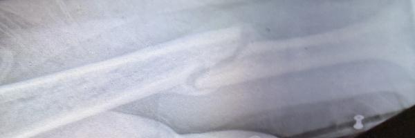
Decollagenified Bones: These were the weakest in this category failing at an average of only 104.2 degrees. Without collagen to provide flexibility to the bones, the bone that was left after we took it out of the oven was extremely brittle. Failure in these bones was fast and apparent, as they would shatter or break very suddenly.
Test 2 bending:
Control Group:Average failure point was 13.6 lbs (with results ranging from 7 lbs to 25 lbs depending on bone mass).Physical Behavior: These bones demonstrated the highest strength of the three groups. However, the researchers noted that determining the exact moment of failure was subjective because the bones were slippery and had a natural curve.Failure Indicators: Failure was identified when the bone showed visible signs of bending or breaking under the sustained hanging weight.
Decalcified Group:Average failure point: Approximately 2.8 lbs.Physical Behavior: While stronger than the decollagenified group, these bones were too flexible to effectively support weight. Researchers observed that the bones lost calcium from the ends inward, often leaving a "harder spot" in the middle.Failure Indicators: Because the calcium providing rigidity was removed, the remaining collagen matrix would flex and sag under relatively low weight compared to the control.
Decollagenified Group:Average failure point: Approximately 0.06 lbs (so small that it appeared near zero on the standard scale).Physical Behavior: These were the weakest bones in the test. Without collagen to provide elasticity, the mineral structure was extremely brittle and required very careful handling to even set up the test.Failure Indicators: Failure was "much easier to tell" than in other groups because it resulted in the bone snapping suddenly and dropping the weights
Test 3 tension:
Control Group: the average failure point for these bones was 45.8 lbs. Some of the bones were very strong. These bones, withstanding up to 55 lbs of weight. Testing was intentionally stopped at 55 lbs because researchers deemed it unsafe to continue adding more weight. The bones were also noted to be very slippery because they were tested immediately after butchering, making it difficult to keep the wires attached.
Decalcified Group: the average failure point was 4.7 lbs. Removing the calcium left only the collagen which made them bendy and weak, which made the bone too flexible to withstand a stretching force.Failure Indicators: These bones snapped much more easily than the control. Researchers identified a consistent weak spot where the bone shaft turns into the joint, which is where the breaks almost always occurred.
Decollagenified Group:Average failure point: Approximately 4.3 lbs.Physical Behavior: Without collagen to provide elasticity and energy absorption, the remaining mineral structure was extremely brittle and fragile.Observations: These bones were so delicate that they were "insanely hard" to even prepare for the test; they would often snap from the slightest tweak while researchers were simply trying to attach the wires before any weight had been applied
Conclusion
Our hypothesis about taking calcium and collagen away was correct. American Journal of Physiology. Cell Physiology. (2020) from our research states that bones with calcium removed become flexible, and bones with collagen removed become brittle.In our experiments we saw similar results to the people who conducted the experiment in American Journal of Physiology. Cell Physiology. (2020).The results of our first experiment showed that the bone with calcium removed broke less quickly under compression. This test was very hard to control perfectly so this might have been incorrect to some degree. but We also think this is because the calcium in the bone provides rigidity and when it was removed the bone was more flexible. The bone with collagen removed broke more quickly under compression compared to the control.The reason that we think this happened is because calcium by itself was too brittle without the collagen for flexibility. Our second test (bending) showed that both collagen and calcium were important for bones to hold up against tension force. Our research found that with some diseases that are caused by a lack of collagen like Osteogenesis Imperfecta make bones extremely brittle and easy to break. In our experiments we found similar results to what our research stated like that the bone with collagen removed snapped more easily because collagen provides elasticity and the bone was too brittle to withstand the tension provided.The decalcified bone showed similar results but for different reasons.Our theory is that when the calcium was removed it left only collagen which made the bone too flexible to withstand the stretch. In our second experiment we also noticed that the bones had a hard spot left at the end after decalcification and in International Osteoporosis Foundation, (2025)we found that there is a hard outer layer and a softer inner layer which shows that the vinegar got rid of most of the outer layer but left a small hard spot still there. In our final experiment we observed similar results to the second experiment; when the calcium and collagen were removed both bones broke more quickly than the control because the bone was either too brittle or too flexible. Probably because the components that we took away provided essential attributes to the bone. Harvard Stem Cell Institute (2020)states that when there isn’t enough calcium in a blood stream the body raids calcium from the bone making it too flexible and we simulated this by taking away the calcium in this experiment. Over all our experiments we found that while all the materials in a bone are needed for its function calcium seems to provide rigidity and collagen provides flexibility.
Application
Real-World Applications
- Understanding bone diseases: Our research helps doctors and scientists better understand conditions like Osteoporosis and Osteogenesis Imperfecta, which make bones weak and more likely to break. By studying bone structure, doctors can develop improved treatments and ways to stop bone deformation before it starts happening.
- Understanding how calcium and collagen affect bone structure and function: Calcium provides toughness and strength to bones, while collagen gives bones flexibility and prevents them from breaking easily. Knowing how these two components work together helps in designing better vitamins, minerals, diets, and medicines to maintain strong bones throughout life.
- Understanding how calcium and collagen impact bone diseases: Many bone diseases occur when there is a problem with low calcium levels or low collagen production. For example, low calcium can contribute to osteoporosis, while low collagen is linked to osteogenesis imperfecta. Studying these connections helps researchers create targeted treatments.
Understanding the importance of calcium and collagen in healthy bones:
Our research shows why proper nutrition and lifestyle choices are important. Eating calcium-rich foods and maintaining healthy collagen consumption through balanced nutrition and exercise can reduce the risk of fractures and bone-related health problems as people get older.
Sources Of Error
uncontrolled/ source of error- secondary effects of vinegar on the bone’s,unable to identify exact moment of bone failure, amount of pressure applied for one turn of the vice handle, how much calcium in one leg bone before test, amount of calcium lost in vinegar, placements of wire on bone, wire not being centerd on bone.
Citations
Works Cited “Bone biology.” International Osteoporosis Foundation, https://www.osteoporosis.foundation/health-professionals/about-osteoporosis/bone-biology. Accessed 19 November 2025. “Bone or cartilage: how stem cells repair bone fractures | Harvard Stem Cell Institute (HSCI).” Harvard Stem Cell Institute\, 27 February 2020\, https://www.hsci.harvard.edu/news/bone-cartilage-van-gastel-2020. Accessed 19 November 2025. Godman, Heidi. “Essential nutrients your body needs for building bone.” Harvard Health, 1 January 2022, https://www.health.harvard.edu/staying-healthy/essential-nutrients-your-body-needs-for-building-bone. Accessed 19 November 2025. “Methods to accelerate fracture healing – a narrative review from a clinical perspective.” Frontiers, https://www.frontiersin.org/journals/immunology/articles/10.3389/fimmu.2024.1384783/full. Accessed 19 November 2025. National Institute of Arthrites and musculoskeletal and skin diseases. “What Is Bone.” What Is Bone, may 2023, https://www.niams.nih.gov/health-topics/what-bone. “Osteoblasts & Osteoclasts: Function, Purpose & Anatomy.” Cleveland Clinic, https://my.clevelandclinic.org/health/body/24871-osteoblasts-and-osteoclasts. Accessed 19 November 2025. “Osteoporosis - Symptoms and causes.” Mayo Clinic, 20 September 2025, https://www.mayoclinic.org/diseases-conditions/osteoporosis/symptoms-causes/syc-20351968. Accessed 20 November 2025. “Osteoporosis: Symptoms, Causes and Treatment.” Cleveland Clinic, https://my.clevelandclinic.org/health/diseases/4443-osteoporosis. Accessed 19 November 2025. steppe, Lena. “Fracture healing research: Recent insight.” December 2023, https://www.sciencedirect.com/science/article/pii/S2352187223000347. NIH. “Physiology, Bone Remodeling.” National Library Of Medicine, National library of medicine, 17 march 2023, https://www.ncbi.nlm.nih.gov/books/NBK499863/. Accessed 22 november 2025. Cellular and Extracellular Matrix of Bone, With Principles of Synthesis and Dependency of Mineral Deposition on Cell Membrane Transport.Schlesinger PH, Blair HC, Beer Stolz D, et al.American Journal of Physiology. Cell Physiology. 2020;318(1):C111-C124. doi:10.1152/ajpcell.00120.2019.Comparison of Different Protocols for Demineralization of Cortical Bone.Pang S, Su FY, Green A, et al.Scientific Reports. 2021;11(1):7012. doi:10.1038/s41598-021-86257-4.The Role of Matrix Composition in the Mechanical Behavior of Bone.Unal M, Creecy A, Nyman JS.Current Osteoporosis Reports. 2018;16(3):205-215. doi:10.1007/s11914-018-0433-0.The Role of Collagen in Bone Strength.Viguet-Carrin S, Garnero P, Delmas PD.Osteoporosis International : A Journal Established as Result of Cooperation Between the European Foundation for Osteoporosis and the National Osteoporosis Foundation of the USA. 2006;17(3):319-36. doi:10.1007/s00198-005-2035-9.The Role of Collagen Organization on the Properties of Bone.Garnero P.Calcified Tissue International. 2015;97(3):229-40. doi:10.1007/s00223-015-9996-2.The Contribution of the Organic Matrix to Bone's Material Properties.Burr DB.Bone. 2002;31(1):8-11. doi:10.1016/s8756-3282(02)00815-3.Type 1 Collagen: Synthesis, Structure and Key Functions in Bone Mineralization.Selvaraj V, Sekaran S, Dhanasekaran A, Warrier S.Differentiation; Research in Biological Diversity. 2024 Mar-Apr;136:100757. doi:10.1016/j.diff.2024.100757.
Reid Bopp and Caleb Fallu declare that we used ChatGPT, an artificial intelligence tool developed by OpenAI, during the early planning stages of our science fair project. ChatGPT was used solely to help brainstorm and narrow down possible research topics and to better understand general background information related to potential subject areas. The tool assisted in generating broad ideas and suggesting directions for exploration. It did not design my experiment, collect data, perform analysis, or write my final report. All research design, hypothesis development, experimental procedures, data collection, analysis, conclusions, and written work submitted for this project are our own original work. Any external sources used for background research were independently verified and properly cited in my bibliography.We understand the importance of academic honesty and affirm that ChatGPT was used only as a preliminary idea-generation tool, in accordance with the policies and ethical guidelines of the Calgary Youth Science Fair. Student Name: Reid Bopp Student Name: Caleb Fallu
Reid Bopp and Caleb Fallu declare that we used NotebookLM, an artificial intelligence research and note-analysis tool developed by Google, during the analysis phase of my science fair project.NotebookLM was used to help organize our notes and identify potential patterns, connections, and themes between my experimental research findings and my background research sources. The tool assisted in highlighting relationships within information that we had already gathered input.NotebookLM did not generate our original experimental data, conduct experiments, determine our conclusions, or write our final report. All experimental design, data collection, analysis decisions, interpretations, conclusions, and written work submitted are our own original work. Any insights suggested by the tool were critically evaluated and verified by me before being included in the project.We understand the importance of academic honesty and affirm that NotebookLM was used only as an organizational and analytical support tool, in accordance with the policies and ethical guidelines of the Calgary Youth Science Fair. Student Name: Reid Bopp Student Name: Caleb Fallu
Acknowledgement
For this science fair project we acknowledge and thank multiple people for helping us to finish our project. These people are David Bopp for helping with conducting my experiments, Janet Summerscales also helped with refining data and our results as well as giving us helpful feedback. We also thank Linsay Stockdale for helping us find credible and informational sources during our background research.