
"Harnessing Stem-Cell Bioengineering": Investigating Chronic Kidney Disease and the Potential of Stem-Cell-Based Artificial Kidneys
Arnav Kangralkar
FFCA South High School Campus
Grade 9
Presentation
No video provided
Problem
Problem 1.1-INSPIRATION Growing up, I’ve been a HUGE hockey fan, and one day was watching a simple hockey game. During this game, unfortunately both goaltenders for the Carolina Hurricanes were injured, and suddenly, there was no one available to tend the net for the rest of the night. However, from the stands came the emergency back up goalie by the name of David Ayres, and he stepped on the ice. Just to let you know, this man had never played a professional game of hockey in his life, and was a Zamboni driver. He proceeded to make 28 saves, and helped Carolina win the game. After this, he became a hero, and was inducted into NHL history. I was inspired by him, and so I decided to do some research about him. Turns out, just prior to his debut, he was dealing with MAJOR kidney issues, and was in critical need of a kidney transplant. Fortunately, his mom was a successful kidney match, and donated her kidney to him.
David Ayres’s story really inspired me, because he was able to return to his career after a huge blockage in his life. Despite all these hardships and complications Ayres has faced, he was able to persist till the end, and eventually became an NHL legend. After thinking about Ayres and his incredible journey to the NHL, it later got me thinking: what if he had never been able to find a kidney donor. In his case, he got lucky with his mother, however over 90,000 Canadians are in need of a kidney, and are patiently waiting. 1.2-RESONSING By studying stem-cell-based kidney bioengineering, I hope to better understand how science can push the boundaries of medicine. Even if the breakthroughs are still developing, every piece of research contributes to a larger effort—a global movement of scientists working toward a future where organ failure does not mean the end of opportunity for a healthy life. What I will be doing Chronic Kidney Disease (CKD) is a progressive disorder affecting over 10% of the global population and is characterized by irreversible nephron loss and declining renal function. Current treatments are limited to dialysis and kidney transplantation; however, donor shortages and long-term complications highlight the urgent need for regenerative alternatives. This project begins with an in-depth examination of kidney anatomy, including both gross and microscopic structures, to understand the functional complexity required for successful tissue engineering. It then investigates the pathological mechanisms of Chronic kidney disease and its global health impact. To address the limitations of current treatments, this study explores stem cell–based bioengineering as a potential regenerative solution. Specifically, it evaluates how Induced pluripotent stem cells (iPSCs) may be directed toward kidney lineage differentiation for artificial tissue development. Additionally, this project incorporates Bioinformatics to analyze kidney gene expression datasets, identify key soluble growth factors and protein signaling pathways involved in nephron development, and determine the optimal molecular microenvironment necessary for engineering functional artificial kidney tissue.
Method
Method In my research study, I will be following a strict guideline of steps. Part 1-The Kidney
- Study The Gross Anatomy of the Kidney
- The the Microbiology of the Kidney
Part 2-Chronic Kidney Disease
- Study the Symptoms of CKD
- Stages of CKD
- Causes of CKD and Complications
- Microbiology
- Epidemiology of CKD
Part 3-Bioengineering A Kidney
- How does Bioengineering a Kidney work?
- 3.2A Driving Kidney Cell Differentiation Using Soluble Factors
- 3.2B Driving Kidney Cell Differentiation Using Soluble Factors
- 3.3-Using the Extracellular Matrix to Drive Kidney Cell Differentiation
- 4.Creating The Best Microenvironment for developing a Kidney
- 5.- Issues with Bioengineering, and why its not feasible
Extra Knowledge This project used a bioinformatics-based research approach to determine the best molecular environment for guiding human induced pluripotent stem cells (hiPSCs) to become nephron cells. Instead of conducting laboratory experiments, I analyzed publicly available scientific databases and peer-reviewed research articles to gather and interpret data. First, I reviewed scientific studies to identify soluble growth factors that researchers have previously used in kidney differentiation experiments. The main factors identified were WNT4, TGFB1, HGF, Activin A, Retinoic Acid, IGF1, PDGFA, and TGFA. I then organized these factors based on their biological roles, such as promoting cell proliferation, directing differentiation, supporting organ development, and regulating epithelial-to-mesenchymal transition (EMT), which is important during kidney formation. Next, I collected gene expression data for selected integrin subunits using the NCBI Gene database. I recorded RPKM expression values for human kidney tissue at 10, 16, and 20 weeks of embryonic development, as well as in adult kidneys. The integrins analyzed included ITGA2, ITGA3, ITGA5, ITGA6, ITGA7, ITGA8, ITGAV, ITGB1, ITGB4, ITGB5, and ITGB8. I then identified which integrins showed the highest expression during embryonic development, since high expression during this stage suggests an important role in nephron formation. These integrins were matched to the extracellular matrix (ECM) proteins they bind to using published interaction data. Finally, I combined the most promising soluble factors and ECM proteins into a proposed differentiation model. Based on my findings, WNT4 supports overall nephron formation, TGFB1 plays a role in podocyte development, and HGF promotes tubular growth. Laminin-511, laminin-521, fibronectin, and vitronectin were identified as the most suitable ECM substrates. Overall, this bioinformatics approach allowed me to logically identify the molecular components needed to recreate a kidney-like developmental microenvironment.
Research
1. Anatomy of the Kidney
The Kidneys are a pair of retroperitoneal organs located between the T12 and L3 vertebrates that consist of an outer and inner medulla, surrounded by tissue and fat. The gross anatomy consists of the following:
- Renal Capsula and Fascia:This part of the kidney surrounds the kidney in a fibrous renal capsule which is surrounded by fat.
- Cortex:This is the outer layer in which the renal corpuscles are located.
- Medulla: The inner region containing renal pyramids separated by renal columns
- Renal Sinus/Pelvis:The central space where urine collects in the major and minor calyces before draining into the ureter
1.1-Internal Structure and Functional Anatomy of the Kidney
The 3 main internal regions in the kidney are called the Renal Cortex, Renal Medulla, and Renal Pelvis. Renal Cortex:The renal cortex is the outermost region of the kidney and is granular(grainy under microscope) due to the presence of nephrons. Also, the cortex provides space for arterioles(small arteries) venules(small veins), and is highly vascular(high blood flow). Also, the hormone Erythropoietin is found in this area. This hormone is necessary for the production of blood cells. Renal Medulla: The medulla is the inner region of the parenchyma of the kidney. This area consists of renal pyramids, which are triangular structures that contain a dense structure of nephrons. Renal Pelvis/Sinus:The renal pelvis contains the hilium. The hilum is the concave part of the bean-shape where blood vessels and nerves enter and exit the kidney; it is also the point of exit for the ureters—the urine-bearing tubes that exit the kidney and empty into the urinary bladder. The renal pelvis connects the kidney to the rest of the body. Renal Arteries: These branch off the abdominal aorta and carry roughly 20-25% of the heart's cardiac output to the kidneys. They divide into smaller vessels, eventually reaching the nephrons for filtration. Renal Veins: These drain the filtered blood from the kidneys and empty into the inferior vena cava. The left renal vein is generally longer, as it must pass in front of the aorta to reach the vena cava, which sits on the right side.
1.2-Microscopic Anatomy:
Nephrons: Nephrons are the microscopic, functional units of the kidney that are responsible for filtering blood, balancing water and electrolytes, and forming urine. Each kidney has over 1 million nephrons! The main task of nephrons is to balance the plasma to homeostatic set points and excrete potential toxins in the urine by doing 3 steps: filtration, reabsorption, and secretion. Components of the Nephron:
- Bowman’s Capsule(Glomerular Capsule): this is the beginning of the nephron that encloses the glomerulus.
- Glomerulus: is a network of tiny blood vessels (capillaries) in the kidney that acts as a primary filter, removing waste, excess water, and small solutes from the blood to initiate urine formation. The glomerulus and Bowman's Capsule together form the renal corpuscle.
- Renal Tubule:A long tube (including the Proximal Tubule, Loop of Henle, and Distal Tubule) that processes the filtrate by reabsorbing water, nutrients, and minerals back into the bloodstream while secreting wastes
- Juxtaglomerular Apparatus (JGA):a specialized structural unit in the kidney located between the vascular pole of the glomerulus and the distal convoluted tubule. It regulates renal blood flow, glomerular filtration rate (GFR), and systemic blood pressure through the secretion of renin and control of sodium
- proximal convoluted tubules (PCTs):most active part of the nephron in reabsorption and secretion, and connects Bowman’s Capsule to The Loop on Henle
- Loop of Henle:The Loop of Henle is a U-shaped segment of the nephron that extends from the proximal tubule into the renal medulla and returns to the distal tubule. It consists of a descending limb, which is highly permeable to water but relatively impermeable to solutes, and an ascending limb, which actively transports Na⁺, K⁺, and Cl⁻ ions
- Distal Convoluted Tubule(DCT): The DCT is essentially the fine-tuner of the nephron — after most bulk reabsorption in the proximal tubule and Loop of Henle, the DCT makes precise adjustments to electrolyte concentrations, pH, and water content.
2.-Chronic Kidney Disease
Before we dive into bioengineering, we need to understand the basics of Chronic Kidney Disease, otherwise known as Chronic Renal Failure. Well, what even is Chronic Kidney Disease? CKD is a long-term condition where the kidneys are damaged(for up to 3 months) and cannot filter blood properly, which allows waste and fluid to build up in the body.
2.1-Symptoms of CKD
CKD symptoms tend to develop slowly over time. As the kidneys are damaged, it causes acids, potassium, and phosphates to build up in the blood. Unfortunately, symptoms for CKD are vague, and many people don’t realize they have CKD until the very last stages. The loss of kidney function can lead to the following symptoms-
- Nausea
- Vomiting
- Loss of Appetite
- Fatigue and weakness
- Sleep problems
- Decrease mental sharpness
- High blood pressure that's hard to manage
- Shortness of breath(if fluid builds up in lungs)
2.2-Stages of CKD
The stages of Chronic Kidney Disease are affected by eGFR. eGFR is the estimated glomerular filtration rate, and estimates how well the kidneys are able to filter waste.
Stage 1: eGFR>90 This means the kidneys have suffered mild damage, and individuals may not have many symptoms. Although, protein may be detected in the urine
Stage 2: eGFR 60-89 This is generally the same as Stage 1, where the kidneys are mildly damaged. They still work well, however traces of protein can be detected in the urine.
Stage 3:eGFR 30-59 This is where individuals have mild to moderate damage done to the kidneys. The kidneys don't work as well as before, and waste can begin to build up in the body can cause severe health complications, such as high blood pressure and bone disease. With treatment and managing a healthy lifestyle, many people in Stage 3 don't move to Stage 4 and 5.
Stage 4:eGFR 15-29 The kidneys don't work as well as before, and are unable to properly filter waste out of the blood and body. This waste buildup in the body can cause severe health complications such as bone disease and heart disease. This is the last stage before complete kidney failure, Stage 5:eGFR <15 This is where the kidneys have completely failed, and there is severe damage to the kidneys. Because the kidneys are unable to filter out waste, waste products build up in the body, and cause severe health complications. Once patients reach this level of kidney failure, the only options for survival are a dialysis or Kidney Transplant.
2.3-Cause of CKD and Complication
Chronic Kidney Disease happens when a disease of the condition hurts the kidney function. The causes of CKD are the following.
- Type 1 or Type 2 Diabetes
- High blood pressure
- Autoimmune disease(such as lupus, sarcoidosis, and Sjogren syndrome)
- Polycystic Kidney Disease
- Prolonged obstruction of the urinary tract.
- Vesicoureteral(Condition that causes urine to back up into kidneys}
Complications are of the following:
- High blood pressure
- Acid buildup
- Fluid retention
- High blood potassium levels
- Anemia
- Heart Disease
- Weak bones
- Damage to central nervous system
- Pericarditis
2.4-Microbiology behind CKD
Chronic Kidney Disease can be caused by many conditions, but the main ones are diabetes and polycystic kidney disease. These are the following steps during CKD
- Initial Nephron Damage:These conditions damaged the glomerulus, tubules, or surrounding blood vessels
- Hyperfiltration(of remaining nephrons) The remaining nephrons work harder, which leads to hyperfiltration. However, this increases pressure inside the glomeruli, which accelerates further damage.
- Progressive Nephrons loss:The damaged nephrons undergo glomerulosclerosis,(scarring of glomeruli) and once a nephron is scarred, it cannot regenerate
- Declining Kidney Function:As more nephrons are lost, the GFR levels decrease, and if severe, can lead to kidney dialysis or transplantation.\
2.5-Epidemiology of CKD
CKD is a global health issue affecting over 10-14% of the population worldwide(800 million people) with prevalence rising due to aging populations and high diabetes/hypertension rates. It is a leading cause of death and disability-adjusted life-years (DALYs), with mortality rates increasing >70% in Canada since 1990.
How it affects Canadians: It affects 1 in 10 Canadians (roughly 4 million people), with the prevalence expected to rise to over 6.2 million by 2050, driven by an aging population and high rates of diabetes and hypertension. It is highly prevalent in rural areas (86.2 per 1000) and disproportionately affects Indigenous populations. Over 50,000 Canadians live with end-stage kidney disease
3.Using Stem-Cells to Bioengineer an Organ
Bioengineering has made meaningful progress in creating and transplanting functional tissues and organs. It focuses on developing methods to engineer biological replacements for damaged or lost tissues and organs. This work is part of the broader and rapidly expanding field of regenerative medicine, which aims to restore or replace impaired body structures. In 2008, researchers achieved a major milestone by performing the first successful transplant of an organ engineered using a patient’s own stem cells—cells capable of developing into different cell types and multiplying to produce more cells. Since then, other tissues and organs, such as urethras, have been bioengineered using a patient’s cells combined with synthetic scaffolds. A synthetic scaffold is a laboratory-made structure designed to support cell growth and tissue development. When synthetic scaffolds are used instead of donor tissue, the need for cadaver donors can potentially be eliminated.
3.1-Stem Cell Bioengineering
Instead of bioengineering an entire kidney, a more feasible goal is to bioengineer a kidney’s nephrons. While commonly used to bioengineer organs, stem cells come in many varieties and some work better in certain situations than others. Some stem cells have been found in the adult kidney, but they are limited. However, embryonic stem cells (or ESCs) and induced pluripotent stem cells (or iPSCs) have been found able to transform, or differentiate, into different kidney cell types. Human embryonic stem cells (hESCs) are isolated from early-stage embryos that have only about 150 cells. Because hESCs are isolated at such an early stage during development, they can multiply and become any cell type in the human body, whether bone, hair, heart, etc. Cells such as hESCs that can give rise to any cell type in the body are referred to as pluripotent
Human induced pluripotent stem cells (hiPSCs)
are adult human cells that have been “reprogrammed” to behave like human embryonic stem cells (hESCs). Like hESCs, hiPSCs are pluripotent and can develop into almost any cell type in the body. In addition, hiPSCs can be patient-specific, meaning a patient’s own cells—such as skin or blood cells—can be collected, reprogrammed into hiPSCs, and then differentiated into the cell types needed, such as kidney cells for bioengineering a new kidney.
To turn hiPSCs into a specific cell type( like kidney cells), researchers often manipulate the cells’ microenvironment so that it mimics the conditions the cells would normally experience in the human body. For example, to produce kidney cells, the cell culture media may include soluble growth factors—or “soluble factors”—that are present during normal kidney development. Similarly, the surface on which the hiPSCs grow can be designed to replicate the structural part of the microenvironment that kidney cells encounter in the body. This surface is often made of proteins from the extracellular matrix (ECM), a mesh-like network that surrounds cells, providing structural support and promoting cell communication. In my science fair project, I will be using online bioinformatics databases to explore how researchers select soluble factors and suitable ECM proteins to guide hiPSCs into differentiating into nephron cells, and how this could ultimately lead to bioengineering a kidney.
How does Bioengineering a Kidney work?
Bioengineering a kidney nephron begins with human induced pluripotent stem cells (hiPSCs), which are stem cells reprogrammed from adult cells and have the ability to become any cell type. First, the hiPSCs are maintained in specialized media to keep them healthy and undifferentiated. They are then guided to become mesoderm cells by activating signaling pathways such as the Wnt pathway, since the kidney develops from the mesoderm during embryonic development. Next, the cells are directed into intermediate mesoderm using carefully timed growth factors like FGF9, and then further specified into nephron progenitor cells, which eventually form nephrons. To support proper differentiation, the cells are cultured on extracellular matrix (ECM) proteins such as laminin, collagen IV, and fibronectin, which replicate the kidney’s natural microenvironment. The cells are then grown in a three-dimensional culture system, allowing them to self-organize into nephron-like structures, including glomerular and tubular components. To improve maturation and functionality, vascularization is promoted using factors such as VEGF to encourage blood vessel formation. In my project, I will use bioinformatics tools and databases to analyze gene expression data and determine which soluble factors and integrins are most suitable for promoting nephron differentiation and artificial kidney development. Although nephron-like structures have been successfully produced in laboratories, creating a fully functional bioengineered kidney remains a major scientific challenge due to its structural complexity and need for proper vascular integration.
3.2A Driving Kidney Cell Differentiation Using Soluble Factors
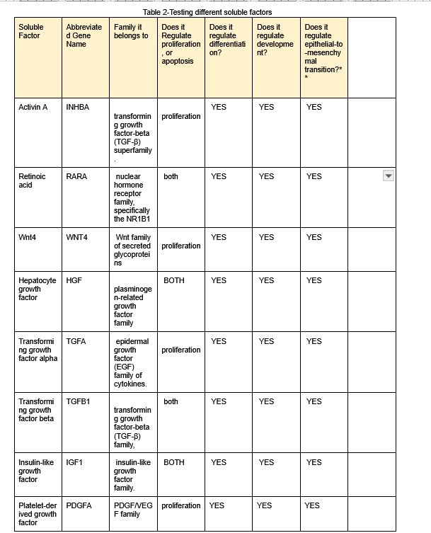
Firstly, I will be using bioinformatics to see which soluble factors would be best to use when trying to differentiate human induced pluripotent stem cells (hiPSCs) into kidney cells, specifically the kidney cells of the nephron.The first table below (Table 1) just summarizes the soluble factors used in key publications of research on turning ECS’s or IPSC’S into different nephron cells.
What the Columns Mean
- Nephron Cell Type → The type of kidney cell the researchers are trying to make.
- Examples:
- Podocytes in the glomerulus → filter cells in the kidney
- Renal tubule cells → tube cells that reabsorb water, salts, etc.
- General kidney development → factors that help the kidney form in general
- Examples:
- Main Supplemented Soluble Factors → The chemicals added to the cell culture to guide stem cells to become that kidney cell type.
- Example: Activin A, Retinoic Acid (RA), HGF, Wnt4, etc.
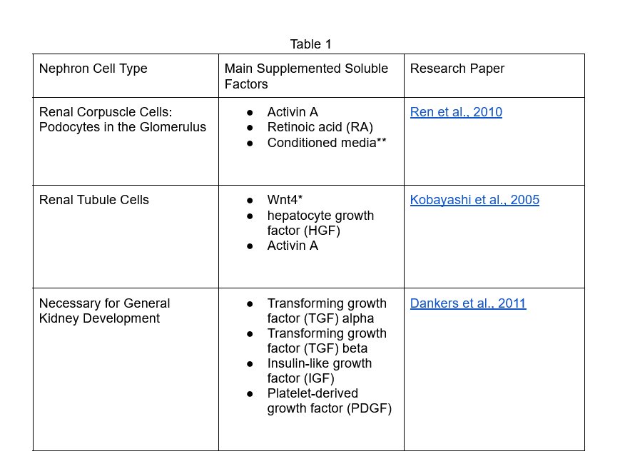
Table 1
| Nephron Cell Type | Main Supplemented Soluble Factors | Research Paper |
|---|---|---|
| Renal Corpuscle Cells: Podocytes in the Glomerulus |
|
|
| Ren et al., 2010 | ||
| Renal Tubule Cells |
|
|
| Kobayashi et al., 2005 | ||
| Necessary for General Kidney Development |
|
|
| Dankers et al., 2011 | ||
3.2B Driving Kidney Cell Differentiation Using Soluble Factors
Next, I decided to use bioinformatics to research and analyze different genes and their properties. Here is each category.
- Soluble factor → The chemical or growth factor (e.g., Activin A, Retinoic Acid).
-
Abbreviated gene name → The shorthand name for that factor in biology databases (e.g., INHBA for Activin A, RARA for Retinoic Acid).
-
Family it belongs to → The type or category of growth factor (like “transforming growth factor” family).
-
Does it regulate proliferation or apoptosis? → Does it help cells grow, divide, or die?
-
Does it regulate differentiation? → Does it tell stem cells to specialize into a particular cell type?
-
Does it regulate development? → Does it help tissues/organs form properly?
-
Does it regulate epithelial-to-mesenchymal transition (EMT)? → EMT is when one type of cell (epithelial) changes into a more flexible type (mesenchymal) — important in kidney development.
This is the Chart
| Soluble Factor | Abbreviated Gene Name | Family it belongs to | Does it Regulate proliferation, or apoptosis | Does it regulate differentiation? | Does it regulate development? | Does it regulate epithelial-to-mesenchymal transition?** |
|---|---|---|---|---|---|---|
| Activin A | INHBA | transforming growth factor-beta (TGF-β) superfamily. | proliferation | YES | YES | YES |
| Retinoic acid | RARA | nuclear hormone receptor family, specifically the NR1B1 | both | YES | YES | YES |
| Wnt4 | WNT4 | Wnt family of secreted glycoproteins | proliferation | YES | YES | YES |
| Hepatocyte growth factor | HGF | plasminogen-related growth factor family | BOTH | YES | YES | YES |
| Transforming growth factor alpha | TGFA | epidermal growth factor (EGF) family of cytokines. | proliferation | YES | YES | YES |
| Transforming growth factor beta | TGFB1 | transforming growth factor-beta (TGF-β) family, | both | YES | YES | YES |
| Insulin-like growth factor | IGF1 | insulin-like growth factor family. | BOTH | YES | YES | YES |
| Platelet-derived growth factor | PDGFA | PDGF/VEGF family | proliferation | YES | YES | YES |
What I noticed: After analyzing the chart, I categorized the genes into 2 different groups. Proliferation:Activin A, WNT4, TGFA, PDGFA. These genes help you grow stem cells so you have enough to work with. They grow a lot of cells. Differentiation:RARA, HGF, TGFB1, IGF1. These genes help guide cells into kidney type cells.(podocytes and tubular cells) EMT: All of them. EMT is important because it forms kidney structures like tubules and glomeruli. After doing some more research on these genes, I was able to make the following conclusions. For the formation of the Nephron as a whole, the gene WNT4 is the best, as it directly signals the cells to make nephron structures. The gene TGFB1 is the best gene for creating podocytes(glomerular cells). And for tubules, the gene HGF is the best.
3.3-Using the Extracellular Matrix to Drive Kidney Cell Differentiation
The extracellular Matrix(ECM) is a mesh-like network of proteins that surrounds cells in the body, giving structural support to the cells and promoting communication among them. Cell receptors are specialized protein molecules located on the surface of, or within, cells that bind to specific signaling molecules. In order for hiPSCs to interact with any ECM, they need specific cell receptors. The ECM is important because it acts like a 3D structural scaffolding. Scientists can study the receptors on real kidney cells to see which ECM they really prefer. So, when bioengineering a kidney’s nephrons, and while they are culturing the hiPSC’s to become kidney cells, the scientists can coat the lab dish with the correct ECM proteins. By doing this, it will help the hiPSCs attach better, survive, and differentiate properly into different cells.
The ECM is a network of proteins that surrounds cells in the body, providing structural support and helping cells communicate with each other. When differentiating hiPSCs into nephron cells, it is important not only to include the correct soluble factors in the culture media, but also to consider the surface the cells are grown on, since the substrate can influence how cells develop. The studies summarized in Table 1 did not compare different substrates. Instead, they first formed clusters of cells called embryoid bodies and cultured them floating in media for two to five days without attaching to a surface. After this floating stage, the cells were placed onto gelatin, a common laboratory substrate made from collagen fragments. However, growing hiPSCs directly on specific ECM proteins may improve how efficiently they differentiate into nephron cells. Understanding which ECM proteins work best could help scientists design better synthetic scaffolds for growing kidney cells that could eventually be used in transplants. In order to attach to ECM proteins such as laminins, collagens, vitronectin, and fibronectin, hiPSCs must have the correct receptors on their cell surfaces.
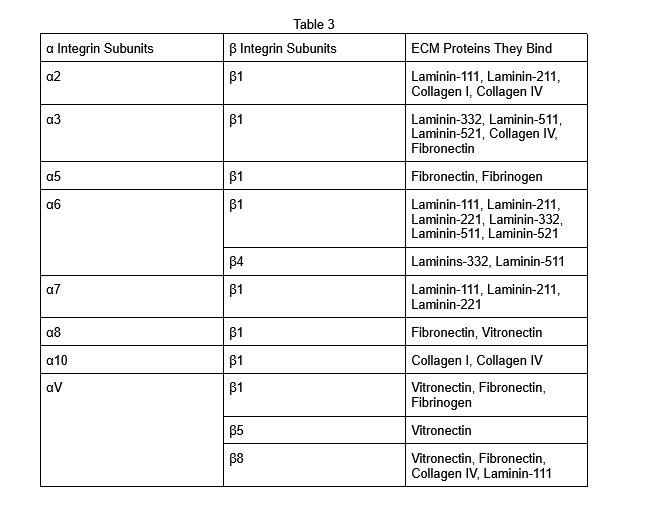
Firstly, before using bioinformatics to collect data on integrin subunits and will be seeing how much it is expressed in an adult kidney, 10w embryo, 16w embryo, and 20w embryo. Because if an integrin is highly expressed during embryonic kidney development, that means it probably plays an important role in forming nephrons. Before I collect data on the integrin subunits, I will be using Table 3 to see all the integrins ( 2 sub units, alpha and beta subunit) and which combinations determine which ECM they build.
| α Integrin Subunits | β Integrin Subunits | ECM Proteins They Bind |
|---|---|---|
| α2 | β1 | Laminin-111, Laminin-211, Collagen I, Collagen IV |
| α3 | β1 | Laminin-332, Laminin-511, Laminin-521, Collagen IV, Fibronectin |
| α5 | β1 | Fibronectin, Fibrinogen |
| α6 | β1 | Laminin-111, Laminin-211, Laminin-221, Laminin-332, Laminin-511, Laminin-521 |
| β4 | Laminins-332, Laminin-511 | |
| α7 | β1 | Laminin-111, Laminin-211, Laminin-221 |
Research Part
Now, I'm going to be using bioinformatics to collect data on all the following integrins. I will be using the website https://www.ncbi.nlm.nih.gov/gene/?term=Homo+sapiens+INHBA to find out information about the integrins, and the RPKM for a 10w embryonic cell, 16w embryonic cell, 20w, and the expression found in the adult kidney. Using these results, I will be able to figure out which integrins have the highest expression in an embryonic kidney. Then, I will connect which ECM proteins to prioritize.
* All numbers stated are the RPKM(reads per kilobase of transcript per million reads mapped) mean.
| Integrin | 10w Embryonic Cell(RPKM) | 16w
Embryonic Cell(RPKM) | 20w
Embryonic Cell(RPKM) | Expression in Adult Kidney |
|
| --------- | ------------------------ | ----------------------- | ----------------------- | -------------------------- | --- |
| a2(ITGA2) | 3.656 | 2.586 | 1.145 | 5.736 ± 1.383 |
|
| a3(ITGA3) | 1.849 | 2.21 | 1.467 | 20.113+3.181 |
|
| a5(ITGA5) | 3.059 | 1.799 | 0.819 | 3.514+0.929 |
|
| a6(ITGA6) | 11.505 | 6.599 | 2.768 | 24.06+4.759 |
|
| a7(ITGA7( | 0.568 | 0.36 | 0.213 | 4.258+0.76 |
|
| a8(ITGA8) | 10.436 | 4.799 | 2.279 | 4.777+0.81 |
|
| aV(ITGAV) | 9.713 | 5.184 | 3.047 | 32.025+0.94 |
|
| b1(ITGB1) | 61.517 | 34.082 | 18.465 | 82.62+6.72 |
|
| b4(ITGB4) | 0.03 | 0.103 | 0.068 | 2.577_1.173 |
|
| b5(ITGB5) | 2.516 | 1.483 | 0.995 | 21.619+2.461 |
|
| b8(ITGB8) | 5.202 | 4.298 | 2.497 | 11.969+2.518 |
|
What I can conclude after my research Top embryonic Kidney Integrins:
| --- |
| ITGB1 |
| ITGA6 |
| ITGA8 |
| ITGAV |
| ITGB8 |
| ITGA2 |
| ITGA5 |
| ITGA3 |
| ITGB5 |
| ITGA7 |
| ITGB4 |
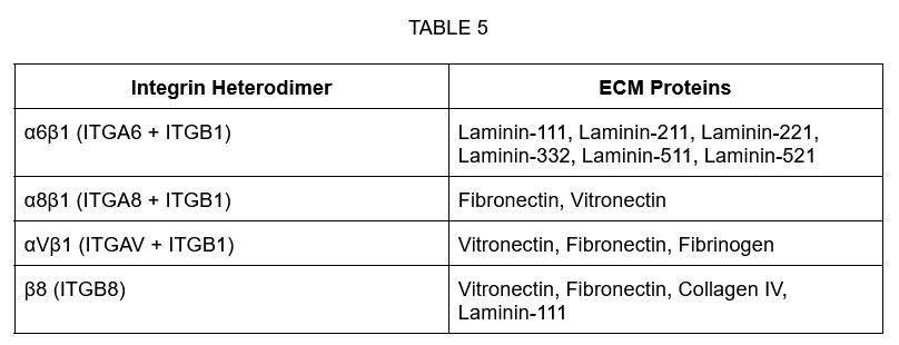
I found out that the integrins with highest expression in embryonic kidneys are ITGB1, ITGA6, ITGA8, ITGAV, and maybe ITGB8. Next, I connected the integrin protein to the ECM proteins it matches to in TABLE 3.
| Integrin Heterodimer | ECM Proteins |
|---|---|
| α6β1 (ITGA6 + ITGB1) | Laminin-111, Laminin-211, Laminin-221, Laminin-332, Laminin-511, Laminin-521 |
| α8β1 (ITGA8 + ITGB1) | Fibronectin, Vitronectin |
| αVβ1 (ITGAV + ITGB1) | Vitronectin, Fibronectin, Fibrinogen |
| β8 (ITGB8) | Vitronectin, Fibronectin, Collagen IV, Laminin-111 |
So the best ECM candidates for differentiating hiPSCs into nephron cells are:
-
Laminins: Laminin-111, Laminin-211, Laminin-221, Laminin-332, Laminin-511, Laminin-521
-
Fibronectin
-
Vitronectin
So, when differentiating a nephrons cells, the best ECm candidates are Laminins: Laminin-111, Laminin-211, Laminin-221, Laminin-332, Laminin-511, Laminin-521, Fibronectin and Vitronectin
4.Creating The Best Microenvironment for developing a Kidney
In order to differentiate hiPSC’s cells into the Kidney’s nephrons, we need to be able to differentiate these kidneys into the right microenvironment. This microenvironment is everything that surrounds the stem cells, which includes the soluble factors and the ECM substrate. Based on my research, I found out the following. Soluble Factors After doing some more research on these genes, I was able to make the following conclusions. For the formation of the Nephron as a whole, the gene WNT4 is the best, as it directly signals the cells to make nephron structures. The gene TGFB1 is the best gene for creating podocytes(glomerular cells). And for tubules, the gene HGF is the best. ECM The integrins with the highest embryonic expression were the following genes. ITGB1, ITGA6 ITGA8 ITGAV. These integrins form these important heterodimers:α6β1 α8β1 αVβ1 And these bind into these ECM proteins, LAMINS, FIBRONECTIN, VITRONECTIN. specific soluble factors play key roles in nephron development. WNT4 is critical for overall nephron formation, as it signals mesenchymal cells to undergo epithelialization and form nephron structures. For glomerular development, TGFB1 plays an important regulatory role in podocyte differentiation and maturation. For tubular formation, HGF is a strong candidate because it promotes epithelial growth and branching morphogenesis, which are essential for forming renal tubules. Together, these factors help recreate the signaling environment necessary for bioengineering a functional nephron. Based on integrin expression data, ITGB1, ITGA6, ITGA8, and ITGAV were highly expressed during embryonic kidney development, particularly at 10 and 16 weeks. These integrins bind laminins, fibronectin, and vitronectin. Therefore, laminin-511 or laminin-521 would likely provide the most supportive substrate for nephron differentiation. Fibronectin may also enhance early differentiation. In combination with appropriate soluble factors that induce mesoderm and intermediate mesoderm formation, these ECM proteins would create a microenvironment that mimics natural kidney development and improves the efficiency of nephron bioengineering. To create the best environment for bioengineering a nephron using sten-cells,
5.- Issues with Bioengineering, and why its not feasible
Structural Complexity
A single kidney contains about one million nephrons, each with:
- A glomerulus (filtration unit)
- Proximal and distal tubules
- Loop of Henle
- Collecting ducts Recreating this precise architecture in a lab is very challenging.
Vascularization (Blood Supply)
Kidneys require a dense network of blood vessels to:
- Filter blood
- Maintain pressure
- Exchange nutrients
Lab-grown kidney organoids often lack proper blood vessel integration, which limits their function.
Functional Maturation
Stem-cell–derived kidney tissues often resemble fetal kidneys, not fully mature adult kidneys. This means:
- Reduced filtration ability
- Incomplete ion transport
- Immature cell types
Scaling the Organ
Growing one nephron-like structure is possible. Growing millions arranged correctly and connecting them to a ureter and blood vessels is much harder.
Risk of Immune Rejection
If cells are not patient-specific, the immune system may attack the bioengineered tissue.
Concerns About Using Stem Cells
Using stem cells is powerful, but there are scientific and ethical concerns:
Tumor Risk
Pluripotent stem cells divide rapidly. If not fully differentiated before transplantation, they may form teratomas (tumors).
Incomplete Differentiation
Not all stem cells turn into the intended cell type. Some may:
- Remain undifferentiated
- Become unwanted cell types
This reduces safety and efficiency.
Ethical Concerns (Mostly with Embryonic Stem Cells)
Embryonic stem cells are derived from early embryos, which raises ethical debates about:
- Moral status of embryos
- Consent and sourcing
High Cost and Technical Complexity
Stem-cell research requires:
- Specialized labs
- Expensive growth factors
- Strict sterile conditions
This limits accessibility.
Genetic Instability
During reprogramming and long-term culture, stem cells can accumulate mutations.
Data
 \
\
Conclusion
In this project, I investigated the optimal conditions for bioengineering a nephron using human induced pluripotent stem cells (hiPSCs). By analyzing integrin expression data, I determined that ITGB1, ITGA6, ITGA8, and ITGAV were highly expressed during embryonic kidney development. These integrins bind extracellular matrix (ECM) proteins such as laminins, fibronectin, and vitronectin, suggesting that laminin-511 or laminin-521 would be ideal substrates for nephron differentiation. Additionally, research on soluble factors revealed that WNT4 is critical for overall nephron formation, TGFB1 plays a regulatory role in podocyte development, and HGF supports renal tubule formation. Together, these ECM proteins and signaling molecules create a microenvironment that mimics natural kidney development. By combining the appropriate substrate and soluble factors in a sequential manner, hiPSCs can be guided toward forming nephron structures, providing a potential strategy for kidney bioengineering and regenerative medicine.
Although this project identified promising ECM proteins and soluble factors, it was based on gene expression data and published research rather than direct experimental testing. Gene expression levels do not always guarantee protein activity, and additional laboratory experiments would be required to confirm the effectiveness of these conditions. Future research could involve testing combinations of laminin-511 and fibronectin with controlled doses of WNT4, TGFB1, and HGF to determine the most efficient differentiation protocol. Further studies could also investigate three-dimensional scaffolds and vascularization, which are necessary for creating a fully functional kidney for transplantation.
I chose to research stem-cell–based bioengineering of kidneys and Chronic Kidney Disease because it represents one of the most powerful intersections of science, hope, and human need. Kidney disease affects millions of people around the world. For many patients, survival depends on dialysis or the hope of receiving a transplant. But donor organs are limited, and far too many people spend years waiting for the chance at a healthy life again. When I learned about this problem, I realized that science has the potential to do more than treat disease—it can transform the way we solve it. Stem cell bioengineering offers a vision of the future where organs could be grown using a patient’s own cells. Imagine a world where someone with kidney failure no longer has to wait on a transplant list, where rejection risks are minimized, and where technology allows us to restore life and health in ways that once seemed impossible. That idea is incredibly powerful to me. What inspires me about this research is not just the biology or the engineering—it’s the human impact behind it. Every advancement in regenerative medicine represents hope for patients and families who are facing some of the hardest challenges of their lives. It reminds us that science is not just about experiments in a lab; it’s about compassion, creativity, and the determination to solve real problems. And this is what bioengineering is all about.
Citations
- Citations https://www.ncbi.nlm.nih.gov/books/NBK482385/
- https://med.libretexts.org/Bookshelves/Anatomy_and_Physiology/Anatomy_and_Physiology_(Boundless)/24%3A__Urinary_System/24.2%3A_The_Kidneys/24.2B%3A_Internal_Anatomy_of_the_Kidneys
- https://courses.lumenlearning.com/suny-dutchess-ap1/chapter/nephrons-structure/
- https://open.oregonstate.education/anatomy2e/chapter/microscopic-anatomy-nephron/
- https://www.sciencebuddies.org/science-fair-projects/project-ideas/BioMed_p013/medical-biotechnology/bioengineer-kidney-with-stem-cells
- https://www.kidneyfund.org/all-about-kidneys/stages-kidney-disease
- https://www.ncbi.nlm.nih.gov/gene/?term=Homo+sapiens+INHBA
-
https://www.ncbi.nlm.nih.gov/gene?Db=gene&Cmd=DetailsSearch&Term=3696#gene-expression
- https://pmc.ncbi.nlm.nih.gov/articles/PMC4373354/
- https://pmc.ncbi.nlm.nih.gov/articles/PMC4373354/
- https://www.uniprot.org/uniprotkb/P26012/entry
- https://share.google/cxmcusmQKkgyEv1n6
- https://www.kidney.org/news-stories/future-artificial-kidneys
- https://www.nature.com/articles/s41581-025-01037-x
- https://wyss.harvard.edu/technology/kidney-engineering-technology-for-new-tissue-replacement-therapies/
- [https://mednews.uw.edu/news/youth-triumphs-test-regenerate-kidney-tissue](https://mednews.uw.edu/news/youth-triumphs-test-regenerate-kidney-tissue%5C)
- https://my.clevelandclinic.org/health/diseases/15096-chronic-kidney-disease
- https://www.mayoclinic.org/diseases-conditions/chronic-kidney-disease/symptoms-causes/syc-20354521
- https://www.ncbi.nlm.nih.gov/search/all/?term=INHBA
- https://www.ncbi.nlm.nih.gov/search/all/?term=rara
- https://www.mayoclinic.org/tests-procedures/bone-marrow-transplant/in-depth/stem-cells/art-20048117
- https://en.wikipedia.org/wiki/Stem_cell
- https://www.ncbi.nlm.nih.gov/books/NBK6259/
- https://cancer.ca/en/cancer-information/cancer-types/wilms-tumour/what-is-wilms-tumour/the-kidneys
- https://my.clevelandclinic.org/health/body/21824-kidney
- https://www.ncbi.nlm.nih.gov/books/NBK482385/
- https://share.google/U2HNjFiE0rCNvFsro
- https://www.diaglobal.org/en/flagship/dia-2026?medium=CPC&source=Google&campaign=23419876784&adgroup=194927575350&content=SearchNetwork&keyword=bio%202026&creative=790890155730&target=&placement=&utm_term=bio%202026&utm_campaign=26101_NA_GAM_Competitor+(gam)&utm_source=adwords&utm_medium=ppc&hsa_acc=6760337270&hsa_cam=23419876784&hsa_grp=194927575350&hsa_ad=790890155730&hsa_src=g&hsa_tgt=kwd-2389104440099&hsa_kw=bio%202026&hsa_mt=b&hsa_net=adwords&hsa_ver=3&gad_source=1&gad_campaignid=23419876784&gclid=CjwKCAiAqprNBhB6EiwAMe3yhlPQzGhHJhbN708b5gHgzhP2vbzp7cF30Pm_1LFZxBQI_NXANZSzMRoC_jQQAvD_BwE
Acknowledgement
I would like to thank my family and friends for helping me and guiding me through this challenging and ambitious project. I seriously wouldn't have been able to finish this project without everyone's support. I would also like to sincerely thank my science fair coordinator, Mr.Nayak on the encouragement throughout this project. And lastly, I'd to appreciate the scientific databases, like NCBI Gene, and the broader scientific community for making research accessible, which made it possible for me to carry out this project entirely through bioinformatics. This project wouldn't have been possible without everyone's encouragement and support, and I'm truly grateful.