
Neurobiologically Informed Schizophrenia Therapeutics: Multi-Modal Characterization and In Vitro Modulation of HERV-K Neuroinflammation
Sara Waqas
STEM Innovation Academy High School
Grade 11
Presentation
No video provided
Problem
Schizophrenia is a chronic neurodevelopmental disorder that affects approximately 24 million people worldwide. It is marked by psychotic symptoms—such as hallucinations and delusions—as well as negative symptoms like reduced emotional expression, motivation, and social engagement. As a result, schizophrenia is among the leading causes of disability globally. Many individuals struggle to maintain employment and relationships, and the condition is linked to a significantly higher risk of suicide: up to 5% of people with schizophrenia die by suicide, and roughly 20% of the homeless population are affected by the disorder. Despite its severity, the biological mechanisms behind schizophrenia are not fully understood. Its recognition as a neurodevelopmental disorder only became widely accepted in the last three decades, making research into how the disorder varies across populations limited. As a result, current treatments often fail to address the disorder’s root causes in favor of temporary symptom suppression, making them largely ineffective. This is evident by the fact that one third of patients remain treatment-resistant.
The current project aims to address the current gap in schizophrenia treatment by working towards neurobiologically precise treatment options. The project extends the neurobiological profiling that was conducted in the 2025 project by introducing a diffusion weighted modality and running hypothesis driven RNA sequencing experiments; designs precise small molecule and RNA-based precise treatment candidates, and finally; validates these targets in-vitro. The aforementioned neurobiological profiling will inform the development of these candidates, with the objective of targeting the core neurobiological deficits that emerge from data of individuals with schizophrenia to promote long term restoration. This research supports the development of precision treatments for schizophrenia that improve long term outcomes, decreasing the debilitating impacts of the disorder and the side effects of antipsychotics on individuals and ultimately improving their quality of life.
Method
Experimental Design:
The project’s experiments will be conducted in three distinct phases.
Phase One - Structural and Hypothesis-Driven Genomic Neurobiological Profiling of Individuals with Schizophrenia and Integrating the Findings of the 2025 Project:
Diffusion Tensor Imaging Analysis:
Materials:
- External Hard-drive
- Andy’s Brain Book
- COINS Database
- MRIcroGL
- SPM
- MRtrix3
- Microsoft Excel
Procedure:
This procedure follows the Diffusion Tensor Imaging Analysis and Fixel Based Analysis procedures outlined in Andy’s Brain Book (Jahn, 2025. doi:10.5281/zenodo.5879293)
Preprocessing:
- Download and extract raw DICOM images from the COINS database onto the external hard-drive
- Upload raw DICOM images into MRIcroGL and use the “convert to NIFTI”
- Combine .bval and .bvec scans in MRtrix using mrconvert
- Use mrview and observe scans in “ortho view” (displays all three angles) to preliminarily check for any striking issues with the data (misalignment, corrupted images, incorrect combination of scans)
- Preprocess data to eliminate warping artifacts by script-running dwi_denoise.
- Check for residuals on scans using mrcalc to ensure brain regions are not disproportionately affected by artifacts.
- Observe scans using mrview and determine if Gibbs’ Ringing Artifacts are present (appear as ripple). If present, remove artifacts by script-running mrdegribbs on affected scans.
- Extract Reverse Phase-Encoded images by converting NIFTI files (the current format of the scans) and add their respective b-values and b-vectors into the headers using mrconvert and mrmath
- Extract b-values from the primary phase-encoded image and combine with the reverse phase-encoded images using mrcat
- Unwarp data and remove eddy currents (final preprocessing step) by script running dwifslpreproc.
- Check the outputted “dwi_post_eddy.eddy_outlier_map” to identify outliers and corrupted slices and remove subjects from study if outlier slices exceed 10% of the total slices present.
- Generate a mask using dwi2mask to ensure the subsequent analysis only evaluates voxels that are part of the brain.
- Use mrview to visually assess if masks were created correctly and align with scans with minimal holes.
- Once these steps are successfully completed, script run the remaining code.
Design and Contrast Matrix for Fixel Based Analysis:
- Create a .txt file with columns of all 1s for equal weighing of all subjects, creating a design matrix.
- Create a .txt file just containing “1” to define the contrast value and average all subject’s fixels.
- Run fibre density, Fibre Cross-Section, (FCS), and Fibre Density and Cross Section (FDC) analysis by running the following in MRtrix:
fixelcfestats fd_smooth/ files.txt *designmatrixfile* *contrastmatrixfile* matrix/ stats_fd/ fixelcfestats log_fc_smooth/ files.txt *designmatrixfile* *contrastmatrixfile* matrix/ stats_log_fc/ fixelcfestats fdc_smooth/ files.txt *designmatrixfile* *contrastmatrixfile* matrix/ stats_fdc/
- Load results by loading the mask into mrview and overlay statistical outputs using Zstat.mif
- Threshold FWE-corrected p-value maps using fwe_1mpvalue.mif at 0.95 to identify fixels considered statistically significant at p < 0.05.
RNA Sequencing Analysis:
Materials:
- NCBI Gene Expression Omnibus
- GALAXY Bioinformatics Web-App
- Microsoft Excel
- DAVID Functional Annotation Web-App
- gProfiler Web-App
- Biotools.fr Web-App
- NCBI Gene Database
- Reactome Web-App
Procedure:
- RNA sequencing raw reads of 10 Bioprojects studying individuals with schizophrenia (Dorsolateral Prefrontal Cortex, Accumbens, Neural progenitors, Astrocytes, Glial progenitors, etc) will be obtained from the NCBI Gene Expression Omnibus.
- In histories for each experiment, use the “Extract in FASTA format” tool to extract raw reads in GALAXY.
- Perform quality assurance analysis using FASTQC to ensure PHRED scores are not <40.
- Perform the “Trimmomatic” operation to omit any reads of low quality.
- Align trimmed reads to the human genome using the HISAT2 alignment tool.
- Quantify gene expression using featureCounts.
- Upload featureCounts “gene counts” file to DESEQ2 for differential gene expression analysis.
- Organize upregulated and downregulated genes and discounted any with an adjusted p-value >0.05 by uploading DESEQ2 results file to Excel.
- Perform pathway enrichment analysis on included genes using DAVID Functional Annotation and identify differentially expressed pathways.
- Congregate results across experiments and identify genes that are differentially expressed across two or more experiments using Excel’s “highlight duplicate” tool.
- Interpret results using existing literature and the NCBI Gene Database.
Data Analysis:
Corrected p-values will be calculated to identify statistically significant fixels. These fixels will be researched in the context of schizophrenia and compared with the fMRI results from the 2025 project to make conclusions regarding the state of structural connectivity of individuals with schizophrenia in order to determine treatment targets. All results will also be verified using existing literature and meta analysis of imaging data to ensure insights are empirically grounded.
Results from RNA-sequencing analysis will be evaluated with the literature to identify unifying themes regarding the neuroinflammation observed in genomic data of individuals with schizophrenia. Genes that are differentially expressed across two or more datasets analyzed will be subject to in-depth research to determine if there is a both a neuroinflammatory mechanism associated with their function and/or if they contribute to a unifying theme of neuroinflammatory disruption through gene enrichment analysis.
Risk Assessment and Mitigation:
No chemical risks arise from the procedure as they will be conducted computationally.
Any participant privacy risks are mitigated because all MRI data is deidentified and essentially stripped of identifying markers during preprocessing, and genomic data utilized is open source, adhering to federal privacy guidelines. No attempts to identify participants will be made.
Ergonomic risks arise from conducting the procedure due to its computational nature and will be mitigated by taking consistent breaks, ensuring proper posture, clearing workspace of anything hazardous, and ensuring the chair and table used are ergonomical.
Phase Two - Therapeutic Design and In-Silico Evaluation/Trialing:
Small Molecule Design and Evaluation:
Materials:
- Chemaxon's Marvin
- SWISSADME
- Tox-Prediction Web App
- ChemMORT
- Pubchem
- AutoDOCK Vina
- PyMOL
- NCBI Protein Bank
- ZINC Database
Procedure:
- Obtain SMILES and IUPAC name of previously constructed molecule from the 2025 project.
- Use insights derived from both literature review and conversations with field experts to optimize the structure of the molecule in Chemaxon’s Marvin.
- Test optimized candidates using Tox-Prediction, SWISSADME, and research arrangements to determine viability.
- Molecularly dock candidates that pass the aforementioned viability tests to three target receptors: 5-HT1A, 5-HT3, and TRKB in AutoDOCK Vina using the following procedure:
- Convert ligand (candidate molecules) into a pdbqt file in AutoDOCK Vina.
- Download target receptors from the NCBI Protein Bank
- Delete water, add polar hydrogens, and Kollman charges to receptors.
- Create grid with locations of ligand and receptor.
- Use command prompt to calculate binding affinity.
- Run tests 5 times and average runs to obtain docking score used to compare viability across candidates and use data from the 2025 project for the three most common antipsychotics to compare effectiveness.
- Find the closest analog of the final treatment candidate using the ZINC database.
Antisense Oligonucleotide (ASO) Design:
Materials:
NCBI Nucleotide Database Pfred ASOptimizer Genscript IDT DNA
- Obtain HERV-K key CDS region from the NCBI Nucleotide Database.
- Input CDS into Pfred app to determine 5 candidate sequences to base ASO on.
- Discuss with field experts on key alterations/optimizers to include in design.
- Input optimized ASO candidate into the ASOptimizer web app and review changes with field experts.
- Test final candidate in IDT’s Oligoanalyzer tool properties page to calculate GC content, melting temperature, molecular weight, extinction coefficient and other measures of ASO stability.
- Generate scramble ASO using Genscript for final candidate and order the scramble and candidate as a “duplexed RNA oligo” from IDT.
Data Analysis:
Candidate molecule’s docking scores, ADME analysis results, and Tox-pro predictions will be compared against each other to determine therapeutic quality.
Candidate ASO’s GC content, melting temperatures, molecular weights, and extinction coefficients will be compared against each other to assess treatment potential.
Risk Assessment and Mitigation:
No chemical risks arise from the procedure as it will be conducted computationally.
No privacy or participant related risks arise from the procedure as there are no participants.
Ergonomic risks arise from conducting the procedure due to its computational nature and will be mitigated by taking consistent breaks, ensuring proper posture, clearing workspace of anything hazardous, and ensuring the chair and table used are ergonomical.
Phase Three - In-vitro Evaluation of Therapeutic Candidates in Primary Rat Culture and HEK293T Cells:
Materials:
- Sprague-Dawley Rat (Strain 400 from Charles River Laboratories)
- HEK293T Cells
- RT-qPCR reagents
- Antisense oligonucleotides
- Eutropoflavin (molecule being tested)
- Calcein-AM and Ethidium Homodimer-1 (cell viability dyes)
- Primary and fluorescently labeled secondary antibodies
- Methanol (used in small quantities as a solvent)
- General laboratory reagents (buffers, media)
- Confocal and fluorescent microscopes
- ImageJ
- RT-qPCR machine
Procedure for Small Molecule Assessment in Primary Rat Culture:
- Prepare primary neuronal culture according to standard laboratory protocol.
- Plate culture into 96 well plate using only the middle 60 wells.
- Place in incubator at 37 °C with 5% CO₂.
- Label this day as DIV 0.
- Maintain cultures with routine media changes until DIV 12.
- Prepare fresh drug stock solution.
- Dilute drug into culture media to obtain the following final concentrations: 20 µmol, 40 µmol, 80µmol)
- Prepare a control condition with no drug (or vehicle only).
- At DIV 12 add drug-containing media to each condition.
- At DIV 14, prepare Calcein-AM and Ethidium Homodimer-1 staining solution according to manufacturer instructions.
- Remove culture media and add staining solution to each chip.
- Incubate cells.
- Rinse gently with buffer if required.
- Image cultures using a fluorescence microscope.
- At DIV 15, remove media and fix cells with fixative solution.
- Wash cultures with buffer, and permeabilize cells.
- Add primary antibodies (Synaptophysin, PSD-95, Neurofilament 160 kDa)
- Incubate.
- Wash cultures to remove unbound primary antibodies.
- Add secondary antibodies (Alexa Fluor 555, Alexa Fluor 633, Alexa Fluor 488)
- Incubate in the dark.
- Wash cultures thoroughly.
- Image using confocal microscopy.
Procedure for ASO Assessment in HEK293T Cells:
Batch 1:
- Plate cells in 96 well plate using only the middle 60 rows.
- Incubate cells at 37 °C with 5% CO₂.
- Allow cells to adhere until reaching around 70% confluency.
- Prepare ASO stock solution.
- Prepare ASO scramble control stock solution.
- Dilute ASO and ASO scramble into culture media to achieve final concentrations of 5 nM, 10 nM, and 20 nM
- Once confluency has been achieved, remove a portion of media from each well.
- Add ASO-containing media to treatment wells.
- Add ASO scramble-containing media to scramble control wells.
- Return cells to incubator.
- At day 3, 48 hours post-treatment, remove media from cells.
- Harvest cells and extract total RNA from cells.
- Quantify RNA concentration and synthesize cDNA from extracted RNA.
- Perform RT-qPCR using validated HERV-K primers (env, pol, and gag) and housekeeping gene (RPL36AL)
- Record Ct values for target genes and housekeeping gene.
Batch 2:
- Repeat steps 1-4.
- At day 3, 48 hours post-treatment, prepare Calcein-AM and Ethidium Homodimer-1 staining solution.
- Remove media and add staining solution.
- Incubate cells.
- Image cells using fluorescence microscopy.
Data Analysis:
For all analysis, statistical significance will be set at p < 0.05.
- Live/Dead Quantification:
Fluorescence images will be analyzed using ImageJ (FIJI) and images will be split into red and green channels. A consistent threshold will be applied across all conditions.
Automated particle analysis will be used to quantify the number of Calcein-positive (live) cells and the number of Ethidium-positive (dead) cells
The live/dead ratio will be calculated for each field of view and averaged across replicates.
2. Immunocytochemistry Analysis
Synaptic density will be quantified by measuring puncta density and integrated fluorescence intensity of Synaptophysin and PSD-95. Colocalization analysis will be performed to assess pre- and postsynaptic alignment. All measurements will be normalized to control conditions.
3. RT-qPCR Analysis:
Ct values from the RT-qPCR instrument will be analyzed by calculating the ΔCt values by normalizing target genes to housekeeping genes and the ΔΔCt values relative to the scramble condition. Finally, ΔΔCt values to relative expression levels using the 2⁻ΔΔCt method and relative gene expression for each ASO dose will be plotted.
Risk Assessment and Mitigation:
Potentially Hazardous Biological Agents:
- Sprague-Dawley Rat Tissue (Strain 400 from Charles River Laboratories.
- HEK293T Cells
Based on the use of cell lines and non-infectious molecular biology techniques, this research is classified as Biosafety Level 2 (BSL-2). Although HERV-K sequences are derived from endogenous retroviral elements, they are not capable of producing infections. Hazardous Chemicals and Devices:
- RT-qPCR reagents
- Antisense oligonucleotides
- Eutropoflavin (molecule being tested)
- Calcein-AM and Ethidium Homodimer-1 (cell viability dyes)
- Primary and fluorescently labeled secondary antibodies
- Methanol (used in small quantities as a solvent)
- General laboratory reagents (buffers, media)
Antisense oligonucleotides, Eutropoflavin, cell viability dyes, primary and secondary antibodies, methanol, and general laboratory reagents will be disposed according to their safety datasheet and/or processed using chemical disposal protocols within the university. Hazardous Activities:
- Handling of mammalian cell cultures
- Pipetting and handling small volumes of chemicals
- Use of a biosafety cabinet
- Use of a PCR machine
- Use of a fluorescent microscope
The in vitro phase involves work with HEK293T cells and primary rat neuronal cultures, as well as the use of antisense oligonucleotides, Eutropoflavin, and standard biology assays which pose a chemical exposure risk. The reagents used when performing RT-qPCR, viability assays, and dissolving Eutropoflavin (methanol) also pose similar exposure risks. Other potential chemical/biological hazards include exposure risks when handling both primary rat neuronal culture and HEK293T cells. As for physical hazards, standard laboratory equipment including centrifuges, PCR machines, fluorescent microscopes, biosafety cabinets, etc will be used in the project and can pose the minor risk of mechanical injury. All biological materials used are non-infectious, and no microorganisms are involved in this study.
All in vitro components of this project will be conducted in a university laboratory under supervision and will follow institutional safety and biosafety guidelines. HEK293T and primary rat cell cultures will be handled using aseptic technique within a biosafety cabinet with laminar flow hoods. In addition to this, standard laboratory personal protective equipment, including lab coats, gloves, and eye protection, will be worn. Moreover, all equipment (centrifuges, PCR machines, fluorescent microscopes, biosafety cabinets) will be used only after proper training. Specifically in the case of the aforementioned reagents, dyes, antisense oligonucleotides, and Eutropoflavin, direct skin and eye contact will be avoided through the use of PPE. All wet lab work will be conducted under the direct supervision of trained and qualified lab members, and the student researcher will be provided all necessary training. Emergency supplies such as eye-wash, and chemical showers are present in the lab space and routinely inspected. Other supplies like spill kits are also present. All work will be conducted in certified laboratory spaces.
Vertebrae Animal Use:
This study will use primary tissue culture extracted from up to 2 pups from the litter of a Sprague-Dawley Rat (Strain 400 from Charles River Laboratories), with the litter being obtained from a separate protocol. In other words, no animals will be euthanized for this project, and tissue was obtained from another approved protocol. Pups will be euthanized by the Qualified Scientist using institutionally approved methods and an ice bath to ensure minimal discomfort and pain. All biological material will be disposed of separately from used glassware and regular waste, and will be processed with biohazard disposal protocols within the university.
While alternative methods for assessing the molecule such as immortalized cell lines and computer-based simulations exist, they are not able to fully replicate the physiological properties of primary neural cells. Moreover, computer-based simulations were used prior to the study to eliminate unpromising candidates to minimize vertebrae use. The use of vertebrates is necessary because primary tissue cultures provide a more biologically accurate model for investigating responses to treatment, especially when studying disorders as complex as schizophrenia. The tissue obtained will be from a litter being used primarily for the host lab’s purpose, meaning no rats will be used for the sole purpose of the project.
The benefits of this research are twofold. First, this research tests the approach of basing treatment candidates on computational results and the integration between the two as a possible approach to drug development. Secondly, this research could contribute to the development of more precise, biologically-informed treatments for schizophrenia that targets the underlying mechanisms of the disorder, which has the potential to catalyze treatment efforts and improve the quality of life of individuals with the disorder.
Research
Results
Diffusion Weighted Magnetic Resonance Imaging (dwMRI) Analysis:
Fourteen white matter tracts demonstrated statistically significant FDC-based differences betweenschizophrenia and control groups. Of these:
- 9 tracts anatomically corresponded to regions identified in the fMRI functional connectivity analysis,demonstrating strong structure–function convergence.
- 12 of the 14 significant tracts showed effects primarily driven by Fibre Density (FD), although Fibre Cross-section (FC) alterations were also present.
Hypothesis Driven RNA Sequencing (RNA-seq) Analysis:
- "Viral process” activation (p = 0.0228), alongside interferon-stimulated and viral RNA sensing pathways.
- BST2 (Tetherin), a canonical retroviral budding restriction factor, was upregulated across datasets(suppresses endogenous retroviral particle release)
- Repetitive element repression and chromatin-silencing pathways were upregulated
- Unfolded protein response and endoplasmic recticulum stress pathways were upregulated, consistent withviral-like protein burden.
- Synaptic, axon development, and receptor tyrosine kinase signaling genes (NTRK2/TrkB) weredownregulated, supporting the 2025 project's GABAergic interneuron hypothesis.
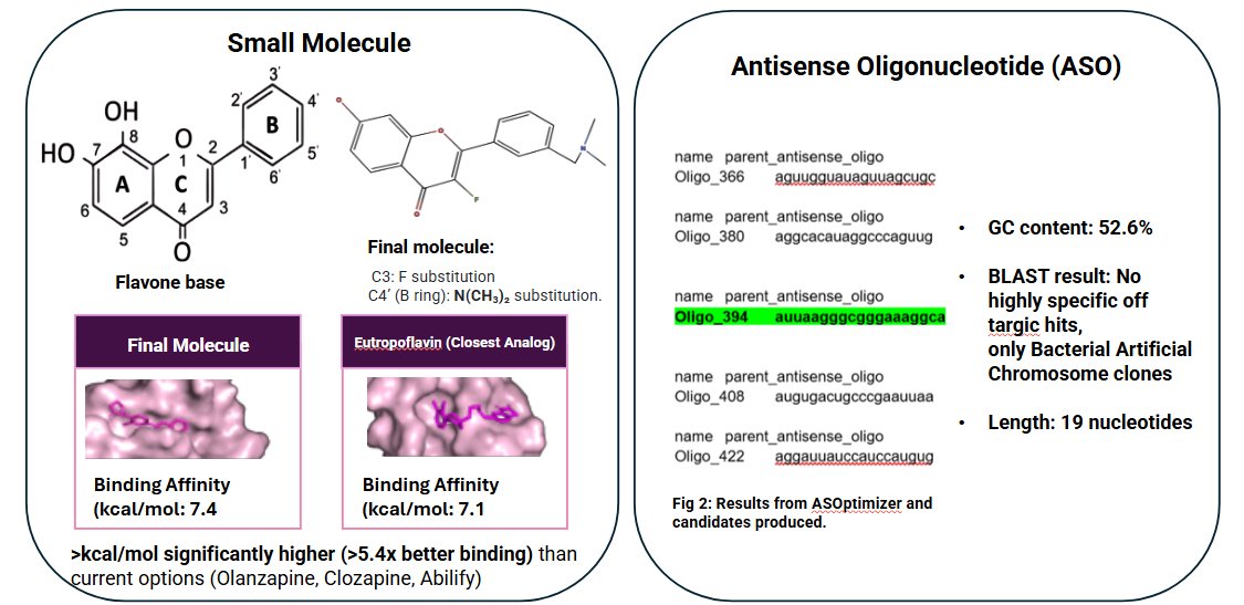
Therapeutic Design Results:
In Vitro Validation:
Small Molecule:
Neurons were treated at 20 µM, 40 µM, and 80 µM. Functional endpoints assessed: • Cell viability (Calcein-AM / Ethidium Homodimer) • Synaptic markers (Synaptophysin, PSD-95) • Neuronal integrity (Neurofilament 160 kDa) Dose-dependent effects quantified relative to vehicle controls.
ASO:
Cells treated with 5 nM, 10 nM, and 20 nM ASO. Primary outcomes: • HERV-K (env, pol, gag) expression using RT-qPCR • Viability assessment (Live/Dead assay) Relative expression calculated using 2⁻ΔΔCt normalization to scramble control.
Data
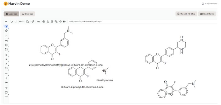 Fig 1: Molecule design in Marvin.
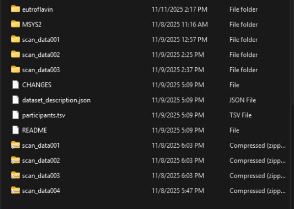 Fig 2: Scan data (dwMRI) used in the project.
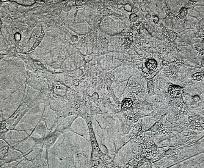 Fig 3: Primary hippocampal neuronal culture on DIV12.
Conclusion
Discussion
Objective 1: Neurobiological Profiling
- Identified cellular vulnerability instead of large-scale anatomical loss in white matter tracts.
- fMRI and dwMRI results were convergent, suggesting structurally compromised tracts are functionally dysregulated.
- Convergent differential gene expression analysis suggested endogenous viral activation, specific drivers pointed to retroviral elements.
Objective 2: Therapeutic Options
- Designed, optimized, and validated a novel candidate in silico.
- Candidate and its closest analog had far superior binding scores (over 5.4x) compared to 3 of the most common antipsychotics:
Clozapine Binding Affinity (kcal/mol): -5.9 Abilify Binding Affinity (kcal/mol): -5.4 Olanzapine Binding Affinity (kcal/mol): -6.4
- Candidate ASO had exceptional properties and no off-target matches, suggesting specific knockdown.
- Avoids indiscriminate inflammatory regulation, targeting drivers.
Objective 3: In Vitro Validation
- Suggests small molecule is not only safe but neuroprotective.
- Large concentrations did not have significant effects on cell proliferation and function.
- Demonstrates safety and pharmacological value of candidate.
- Reductions in HERV-K env and gag amount without significant effects on housekeeping gene demonstrates knockdown and precision.
Conclusion
A Precision Approach Moves Away From Symptomatic Treatment To Targeting Key Drivers
- White matter disruption appears driven by axonalmicrostructural vulnerability rather than gross anatomicalloss and converges with fMRI results.
- Convergent transcriptomic findings implicate endogenousretroviral activation as a potential upstream driver ofinflammation.
- Targeted therapeutic design and in vitro validationdemonstrates the feasibility of precision intervention.
- This work supports a shift toward biologically stratified,mechanism-driven treatment development in psychiatry.
Citations
Only sources used for the construction of this portal were included.
Andrade, C. (2020, January 6). Sample size and its importance in research. Indian journal of psychological medicine. https://pmc.ncbi.nlm.nih.gov/articles/PMC6970301/ Borrego-Ruiz, A., & Borrego, J. J. (2025, March 10). Involvement of virus infections and antiviral agents in schizophrenia. Psychological medicine. https://pmc.ncbi.nlm.nih.gov/articles/PMC12055031/
Dhuri, K., Bechtold, C., Quijano, E., Pham, H., Gupta, A., Vikram, A., & Bahal, R. (2020, June 26). Antisense oligonucleotides: An emerging area in drug discovery and development. Journal of clinical medicine. https://pmc.ncbi.nlm.nih.gov/articles/PMC7355792/ Hubrecht, R. C., & Carter, E. (2019, September 30). The 3Rs and humane experimental technique: Implementing change. Animals : an open access journal from MDPI. https://pmc.ncbi.nlm.nih.gov/articles/PMC6826930/
Kim E;Carreira Figueiredo I;Simmons C;Randall K;Rojo Gonzalez L;Wood T;Ranieri B;Sureda-Gibert P;Howes O;Pariante C;Nima Consortium None;Pasternak O;Dell’Acqua F;Turkheimer F;Cash D; (n.d.). Mapping acute neuroinflammation in vivo with diffusion-MRI in rats given a systemic lipopolysaccharide challenge. Brain, behavior, and immunity. https://pubmed.ncbi.nlm.nih.gov/37482203/
Klein, H. C. (2017a, December 13). Silencing of viral elements: An available cure for schizophrenia?. Frontiers in psychiatry. https://pmc.ncbi.nlm.nih.gov/articles/PMC5733551/
Kotsiri, I., Resta, P., Spyrantis, A., Panotopoulos, C., Chaniotis, D., Beloukas, A., & Magiorkinis, E. (2023, June 9). Viral infections and schizophrenia: A comprehensive review. Viruses. https://pmc.ncbi.nlm.nih.gov/articles/PMC10302918/
Liu, X., Chan, C.-B., Jang, S.-W., Pradoldej, S., Huang, J., He, K., Phun, L. H., France, S., Xiao, G., Jia, Y., Luo, H. R., & Ye, K. (2010, December 9). A synthetic 7,8-dihydroxyflavone derivative promotes neurogenesis and exhibits potent antidepressant effect. Journal of medicinal chemistry. https://pmc.ncbi.nlm.nih.gov/articles/PMC3150605/
Plank JR;Morgan CA;Dell’Acqua F;Sundram F;Hoeh NR;Muthukumaraswamy S;Lin JC; (n.d.). Mapping neuroinflammation with diffusion-weighted magnetic resonance imaging: A randomized crossover study. Biological psychiatry. Cognitive neuroscience and neuroimaging. https://pubmed.ncbi.nlm.nih.gov/40379248/ Rinaldi, C., & Wood, M. J. A. (2017, December 1). Antisense oligonucleotides: The next frontier for treatment of neurological disorders. Nature News. https://www.nature.com/articles/nrneurol.2017.148
Acknowledgement
Thank you to:
The Naweed Syed Lab, including Dr. Syed, Zainab Khan, Fahad Iqbal, Badra Abbas, and all other members, for their support in the in vitro validation portion of the project. Your constant support and mentorship were invaluable.
Dr. MacEachern at the Precision Neurodevelopment Lab, for being an incredible mentor, PI, and support, and providing the inspiration for the project.
The European Commission, for their award providing funding for the project.
My friends, teachers, and supporters!
Individuals at the COBRE institute for championing open access data.
And finally, and most pivotally, individuals with lived experience and schizophrenia for both participating in the studies that I have referenced in my project as well as talking to me throughout this project and sharing your invaluable experience. Thank you for being so open to my work and advancing science research in general.