
Is it Possible to Identify and Stimulate the Firing Patterns of Neurons Related to a Specific Memory and use this as an Alzheimer's Therapy?
Jonah Lam, Kaiden Manji
FFCA High School Campus
Grade 9
Presentation
No video provided
Problem
Who are you? This is a question that so many families have to witness their loved ones say after they get diagnosed with dementia. In the world its estimated that 55 million have dementia with 70% of these cases being Alzheimer's. According to the World Health Organization (WHO) dementia is the 7th leading cause of death and is one of the key contributors to disabilities in elder people. Even worse is that the illness only gets worse over time and it leaves many peoples loved ones to take care of them 24/7 or send them off to a nursing home. In simple terms dementia is a disease that destroy nerve cells which if Alzheimer's can take away a persons memory. The entorhinal cortex is like a bridge to the hippocampus which is key in making new memories. The entorhinal cortex can also help in the process of retrieving memories which is why it is so important. It is often one of the first parts in the brain to deteriorate when a person has Alzheimer's and it's why it could be the key to a cure. Scientist at LSU are currently researching what they call the "poorly understood entorhinal cortex". If we could learn more about the entorhinal lobe the possiblities are endless.
Method
Our Hypothesis/Question we want to answer at the end of our research is....... Is there any way that we can restore memory from people who have Alzheimer's by studying the neurons in the entorhinal cortex?
Research
To try and figure out how the entorhinal cortex works and what it has to do with memory and memory loss we first have to understand what dementia is, how it is caused and map out how neurons work specifically in the entorhinal lobe. Dementia is an overarching name for types of memory loss with the most common type of dementia being Alzheimer's which is more common in elderly people. Some common symptoms of Alzheimer's disease are repeating questions over and over, forgetting what task you are doing, misplacing items and overtime forgetting the names of family members. The exact cause of dementia is still not clearly known however scientist are working hard everyday to further develop more research on the rather blurry topic. What is currently believed is that genetics and your environment are what play a factor overtime in your mental state. A percentage of the people who get dementia are classified as specifically genetic dementia where they have a genetic modified that almost guarantees that they will get dementia as a middle aged person. Alzheimer's specifically is when brain proteins don't work properly causing neurons to get damaged and die overtime. This process actually starts way before the first symptoms are shown. Dementia first attacks the memory parts of the brain however then it spreads and by the end the brain usually shrinks in size. Scientist have two reason for the deterioration of the brain. One being fragments of protein called Beta-amyloid clump together forming plaques that interfere with the brain. Secondly, tau proteins alter the shape of brain cells, which in turn, block essential nutrients from reaching the brain. These two combined can damage and kill cells which is basically what dementia is as a whole. Some factors that affect Alzheimer's are your age, genetics/family background and lifestyle. For example the Alzheimer's society states that 2% of people have dementia from ages 65-69 while 33% have a type of dementia ages 90+. Women have a higher chance to be diagnosed with dementia more than men because they tend to live longer lives. There are ways to slow or potentially stop dementia by changing your lifestyle. Doing things like exercising everyday and having a healthy diet can reduce the risk of dementia at an older age. The brain is one of the most complex organs in the body that controls the nervous system. Neurons are specialized cells in the brain that transmit electrical signals across the brain. There are approximately 1 billion neurons in the human brain which all serve an important purpose. There are three main parts of the brain the cerebellum, cerebrum and brain stem. They all serve unique purposes.The cerebrum is the biggest part of the brain and runs your thoughts, emotions, speech and sensory emotions. The entorhinal cortex is part of the cerebrum though it is located deep in the temporal lobe. The cerebellum is responsible for coordinating involuntary movement, balance and posture. The brain stem connects the cerebrum to the spinal cord and controls functions like breathing, heart rate, blood pressure and sleep patterns. To try and figure out how studying neurons can fix Alzheimer's we first must know what neurons actually are, what they do and how they are damaged. We also researched specific ways to fix or excite neurons which can reverse loss of memories. Our body has lots of specialized cells the main one in the brain is known as neurons. Neurons are specialized cells in the brain that transmit electrical and chemical signals . The human brain relies on neurons as they are key to biological communication. The branches on neurons are called dendrites. These dendrites are the part of the cell that receives chemical signals and then turns them into electrical signals which are processed inside the cell body. If the impulses are powerful enough an electrical action potential travels down the insulated axon and toward the terminal button This whole process leads to the release of neurotransmitters which in turn allow messages to travel from one cell to another. When somebody has dementia these cells become damaged which messes up the whole communication process. As we talked about earlier beta-amyloid protein clump together causing plaques that interfere with the brain and tau alters the brain if you have some types of dementia including Alzheimer's. This causes neurons to lose communication between dendrites and in turn disrupts the brains process of thinking. Overtimes neurons completely die off and from there it is even harder to repair. after some research we found that there may be some ways to fix this problem by trying to excite the neurons before they die off however this is still unknown and a long stretch. Place cells are a very important part of this project. Place cells are a type of cell that help you understand where you are in space. They are located in the hippocampus which neighbours the etorhinal cortex. Each cell activates when you are in a specific place. For example if you walk by your school that place cell would fire off.Together thousands of these cells map out the area around you creating spatial awareness. Place cells connect memories to places and navigate through places you have been to many times. When someone has Alzheimer's they often lose neurons in the hippocampus first which is why they can get lost sometimes. Why place cells are important is by studying there patterns we can try to find ways to activate these cells which could lead back to a recovery of certain kinds of memories. The hippocampus which is a part of the brain that is essential for short term memory and long term storage. The hippocampus contains place cells, which fire when you are in a specific location that create memories for specific places.
The Columbia Biomedical Experiment on Memory Trace Cells
In 2019, a team led by Columbia Engineering neuroengineers found evidence that the individual neurons in the human brain target specific memories while recalling them. The experiment was simple. Neurosurgical patients with electrodes already implanted into their brains were put through a task in which they had to locate and object in virtual-reality (VR). The researchers then studied how a patient's brain signals corresponded to their behaviour while performing the task. In this VR task, patients were asked to explore the virtual space using laptops or handheld controllers. While exploring, subjects would learn the locations of four different objects. Once all objects had been located by the subjects, researchers removed them and asked subjects to navigate through the same environment and locate, as well as mark, the position of one of the four objects for each trial. The team of researchers measured, recorded and analyzed the activity of neurons during each subject’s trial. They found specifically tuned spatial neurons that are similar to “place cells” or “grid cells” (specialized neurons that only fire when a human or animal is in a specific, localized area of an environment, acting as an internal GPS) that fire when patients moved through certain spaces. These neurons would fire no matter what the patient's target object was. Contrary to these neurons, there were also other neurons that activated only in locations that were related to the object the patient was recalling in that trial. If the subjects were told to switch the object they were locating, the neurons changed their behavior to match the new object they were remembering. Researchers also discovered that they could decipher which object a patient was remembering based on the activity of these neurons. “Our study demonstrates that neurons in the human brain track the experiences we are willfully recalling, and can change their activity patterns to differentiate between memories. They’re just like the pins on your Google map that mark the locations you remember for important events,” Salman E. Qasim, PhD student and lead author of the study says. “This discovery might provide a potential mechanism for our ability to selectively call upon different experiences from the past and highlights how these memories may influence our brain’s spatial map.”
National Institutes of Health (NIH) Firing of Neurons during Memory Recollection Research
A lot of different factors can play into the process of triggering a memory, including sights, smells and sounds that are familiar. Previous studies have suggested that, in rodents, memories are stored as specific and unique firing patterns of neurons. These patterns are repeated every time a memory is triggered. A research team led by the NIH’s (National Institute of Health) National Institute of Neurological Disorders and Stroke’s (NINDS) Dr. Kareem Zaghloul conducted a study to discover whether a similar activity in the brain happens when humans recall memories. The subjects for this study were patients with epilepsy, and whose seizures weren’t able to be controlled with drugs. To aid with this experiment, patients' brains were temporarily embedded with electrodes, allowing scientists to measure neural activity and monitor how memories are stored as well as recalled. The six patients involved in the study were sat in front of a screen and told to learn a pair of two completely unrelated words. While the subjects underwent this task, the researchers monitored the activity of thousands of individual neurons. The brain regions scientists specifically focused on were the anterior temporal lobe (a brain language center) and the medial temporal lobe (the area linked to memory recollection in rodents). They also looked at high frequency brain activity that occurs when multiple neurons are activated at once, a process called “ripples”. These ripples occur in the brain just a fraction of a second before a memory is recalled. When subjects learned these word pairings, unique firing patterns of individual neurons emerged. Later in the trial, the patients were shown one of the two words in the pairing. When they recalled the second word, an extremely similar (but not identical) firing pattern occurred in their brains just a fraction of a second before they remembered the second word. Scientists also discovered a link between the ripples and the firing patterns in the brain. The ripples occurred in the medial temporal lobe just milliseconds before the firing patterns did in the anterior temporal lobe while the patients were learning the words. This discovery proposes that memories are stored in or involve coordinated replay of neuronal firing patterns in the human brain.
External Excitation of Neurons using Electric and Magnetic Fields in One and Two Dimensional Cultures - NIH
A neuron fires action potential when its membrane potential exceeds a certain amount. Typically, this is a result of chemical inputs to a neuron's synapses. However, neurons may also be excited by an imposed electric field. Recent clinical applications use external electric fields to activate these neurons. Here, some techniques used for the controlled application of an external electric field on neurons are explained. These techniques could be one or two dimensional. 2D cultures don't have preferred orientation, and their simulation is isotropic. It does not depend on the orientation of the field applied. When the 2D field is rotating, shorter durations are required. This is the opposite of 1D cultures, which are extremely dependent on the orientation of the field. When the field is oriented with the pattern, the duration of a fixed amplitude field needed is much shorter than if the field was perpendicularly oriented. If the duration is kept constant and the field and pattern are parallel, then the field needed to excite the culture is a lot smaller. When oriented perpendicular to the pattern, the pulse needs to be much longer, and even then dendrites are what are mainly stimulated. When electric fields, whether they may be directed or induced by a magnetic field, are used for neuronal stimulation, this is extremely important. If the area of the brain that is targeted has bundles of axons, these axons can be excited by orienting the field in their direction and then need less power or a shorter duration of the field for stimulation. A rotating field is more efficient if the brain region does not have preferred orientation of the field. Longer pulses are more effective if dendrites are the stimulation target. The use of magnetic fields for neuron stimulation are for when a magnetic pulse induces an electric field that excites neurons. Magnetic fields are used in the stimulation of axons rather than dendrites, as currently achievable magnetic pulse durations come in microseconds, while dendrites response times are in milliseconds. The response of axons is maximized when they are parallel to the electric field and is diminished when they are perpendicular. This means, when using magnetic excitation, we should strive to get the induced electric field parallel to the targeted axons.
Data
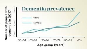 This is a graph showing the number of people that have dementia based off of gender and age. This graph is from the Queensland Brain Institute.
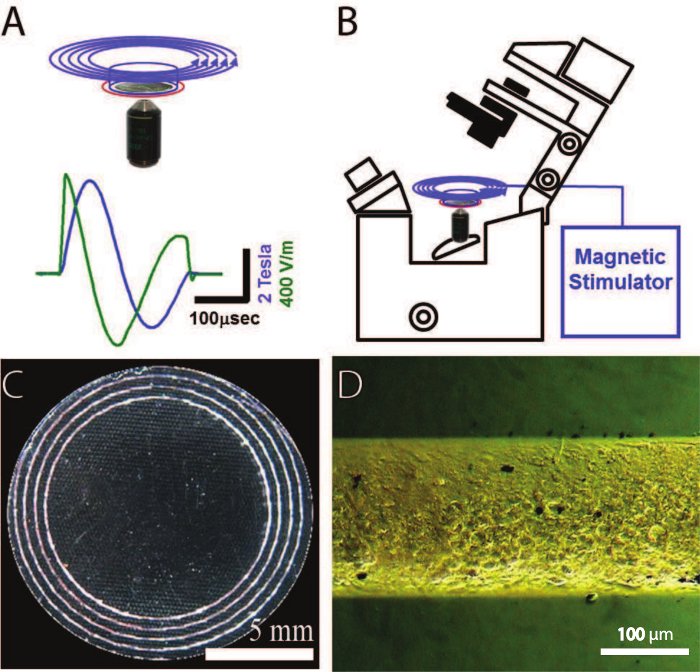 This is a photo exemplifying how to use magnetic feilds and orient them for neuron exitiation.
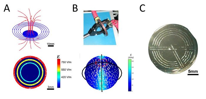 This photo shows the orientation and calculated
Conclusion
To conclude, the brain is a very complex system in the brain. It is even more complex to try to solve Alzheimer's and study complex neuron patterns. All of our research is very theoretical and still in being tested could damage the brain further and we cannot test our solutions on actual people. Though we found one possible way to excite the neurons in the brain we don't know if this would actually work though with the correct technologies it may be possible. It is not widely available or commonly researched right now as Alzheimer's as a whole is something that neurologists are still fully trying to understand. Overall, across the whole time period we researched how the brain works, what dementia is and how it impacts daily life. We then studied patterns in the brain while also learning what neurons are and how they function. We then looked far and wide for any possible solutions to try and potentially gain back memories. We did find that there may be a way to solve our problem by using magnets to excite neurons and cause firing patterns which would in turn lead to recovery of memories. It is important to mention that this discovery is still largely theoretical and in the works. The goal of our research was to answer our research question/hypothesis. We wanted to find out if there was a way to restore memory in dementia patients by studying the entorhinal cortex. I feel like we did a pretty good job at this as we looked at complex studies to come to a discover that there may be a way even though it is a blurry one.
Citations
References
Cancer Research UK. (2020). What are the different parts of the brain and what do they do? | Cancer Research UK. In YouTube. https://www.youtube.com/watch?v=iomhlXlisKI Entorhinal Cortex - an overview | ScienceDirect Topics. (2016). Sciencedirect.com. https://www.sciencedirect.com/topics/neuroscience/entorhinal-cortex Johns Hopkins Medicine. (2025). Brain Anatomy and How the Brain Works. Johns Hopkins Medicine; Johns Hopkins Medicine. https://www.hopkinsmedicine.org/health/conditions-and-diseases/anatomy-of-the-brain Smile and Learn - English. (2020). The Brain for Kids - What is the brain and how does it work? In YouTube. https://www.youtube.com/watch?v=c9HK59FaoMI
References
Entorhinal Cortex - an overview | ScienceDirect Topics. (2016). Sciencedirect.com. https://www.sciencedirect.com/topics/neuroscience/entorhinal-cortex Medvedeva, V. P., & Pierani, A. (2020). How Do Electric Fields Coordinate Neuronal Migration and Maturation in the Developing Cortex? Frontiers in Cell and Developmental Biology, 8. https://doi.org/10.3389/fcell.2020.580657 Memories involve replay of neural firing patterns. (2020, March 16). National Institutes of Health (NIH). https://www.nih.gov/news-events/nih-research-matters/memories-involve-replay-neural-firing-patterns New study uncovers how the brain revises memories. (2025, July 15). Department of Psychology. https://www.psych.utoronto.ca/news/new-study-uncovers-how-brain-revises-memories Specific Neurons that Map Memories Now Identified in the Human Brain. (2019, November 8). Biomedical Engineering. https://www.bme.columbia.edu/news/joshua-jacobs-neurons-map-memories Stern, S., Rotem, A., Burnishev, Y., Weinreb, E., & Moses, E. (2017). External Excitation of Neurons Using Electric and Magnetic Fields in One- and Two-dimensional Cultures. Journal of Visualized Experiments : JoVE, 123, 54357. https://doi.org/10.3791/54357
Acknowledgement
We would like to acknowledge our parents for the support along the way as well as paying the fee. We would also like to acknowledge our teacher Ms Fan for helping us along the way with ideas and questions. We'd also like to thank the bus drivers for allowing us to carry our tri-fold on the bus.