
How Can AI Help with the Accurate Detection of Eye Cancer?
Grade 9
Presentation
No video provided
Problem
Eye cancer is the development of cancerous tumors in or around the eye that can grow and destroy nearby tissue. The most common types of eye cancer are melanoma, lymphoma, retinoblastoma. Currently an accurate diagnosis of eye cancer requires a thorough proedure of the patient going through multiple examinations, and scans. Furthermore the detection of a tumor isnt necesarily always a cancerous tumor, which potentialy leave the pataient to ensue further diagnosis. All of these examinations and scans require time and money, which the patient may or may not have.
Method
So what exactly are the current methods of diagnosis? Majority of eye cancer diagnosis' are done to find out if the patient has a tumor or the size of a tumor.
1. Eye Exam
An eye exam for melanoma often comes with a healthcare profesional examining the eye, looking for blood vessels that may be larger than normal. The second part of the eye exam involves looking inside the eye often using a bright light worn on the health care specialist's head.
2. Fundus Photography
A test that that takes coloured pictures of the inside surface of the eye, known as the fundus. The fundus contains the retina, optic disc, macula and blood vessels. These pictures of the fundus can be used to detect eye melanoma along with tracking melanoma over time.
3. Eye Ultrasound
Uses high frequency sound waves to create images of the eye. The sound waves come from a device called a transducer, which is placed on the patient's closed eye lid or front surface of the eye.
4. Eye Angiography
A test that creates pictures of blood vessels in the eye, by injecting a coloured dye into a vein in the patient's arm. As the dye travel's to the patients eye, a camera with special filters, that detect the dye, takes several pictures over the span of a few minutes.
5. Optical Coherence Tomography
Utilizes light waves to make pictures of the eye, specifically the uvea and the retina both of which may show an eye melanoma.
6. Eye Melanoma Biopsy
Removing a sample of tissue for lab testing. Biopsy is rarely needed for the diagnosis, however it is still used in specific situations.
Now what benifits does AI bring to the table?
Anil Parwani, director of the Division of Anatomical Pathology at The Ohio State University Comprehensive Cancer Center - Arther G. James Cancer Hospital and Richard J. Solove Reasearch Institute states that we are nowhere near unsupervised AI diagnosis, however they are still powerful a powerful tool for pathologists to confirm diagnoses. Anil Parwani also says AI can be used to locate concerning features outside of the cancer that are not obvious to the naked human eye. These details can help create a list of patients ranking from high to low risk cases, allowing for those in dire need of help to recieve it at a swifter pace as compared to before. As of now AI is unable to make an accurate diagnosis on its own however once a pathologist has made the diagnosis, AI can assist in finding aditional clues that will assist the treating oncologist, a medical professional quallified to to diagnose and treat tumors, to come up with a personal treatment plan for the patient, which is even more helpful when dealing with rare cancers. “This is a very powerful technology, and we should be very cautious about how we harness it in medicine to improve care for our patients. It is an incredible diagnostic support and validation tool worthy of continued evaluation,” he said.
Research
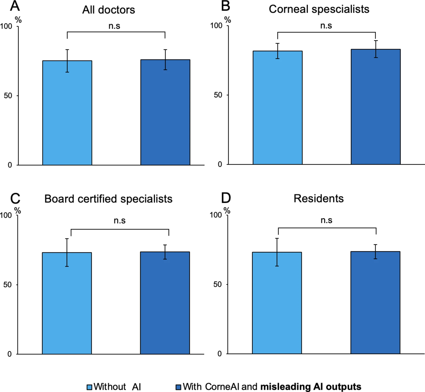
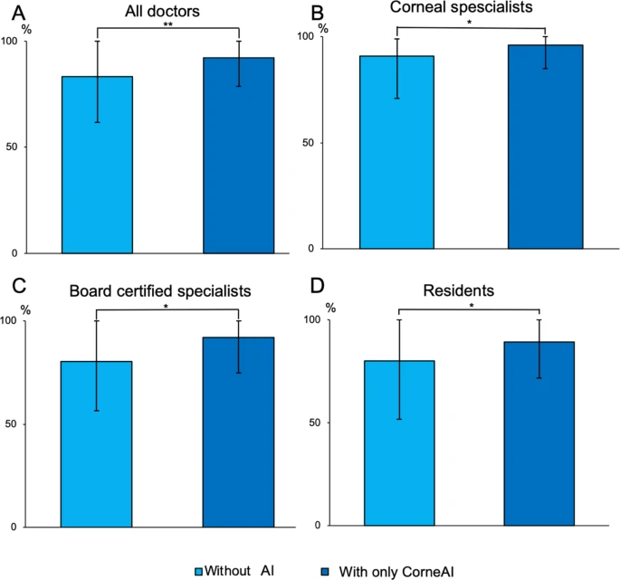
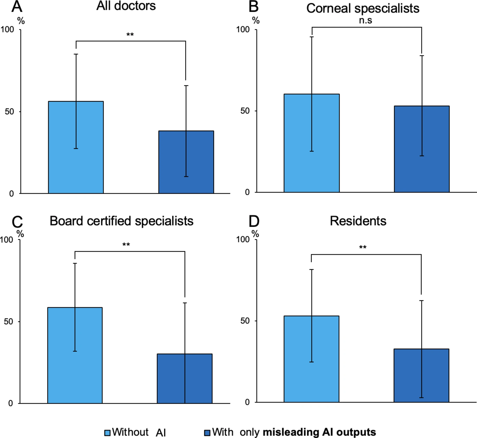
In a study done by Japanese researchers published on January 9th, 2025, an AI, known as CorenAI, was created with the sole purpose of classifying images in the eye that were indicative of corneal diseases. The study included 7 corneal specialists and 16 non-corneal-specialist ophthalmologists all of which were initally only provided images and then the AI's results. However the AI's results were altered to 70% correct answers and 30% incorrect answers. The general accuracy of the ophthalmologists did not change very much once the correct AI results were provided, with an accuracy of 75.2% before and 75.9% after. The biggeer change was seen when the AI provided incorrect information. The corneal specialists had an accuracy of 60.3% before and an accuracy of 53.2% after the false information was provided. However the non-corneal-specialists witnessed a significantly greater drop in their accuracy going from 54.5% to 31.6% after the influence of the AI.
This research was conducted by Hiroki Maehara, Yuta Ueno, Takefumi Yamaguchi, Yoshiyuki Kitaguchi, Dai Miyazaki, Ryohei Nejima, Takenori Inomata, Naoko Kato, Tai-ichiro Chikama, Jun Ominato, Tatsuya Yunoki, Kinya Tsubota, Masahiro Oda, Manabu Suzutani, Tetsuju Sekiryu & Tetsuro Oshika,
| No AI Round #1 | Correct AI Information | No AI Round #2 | Incorrect AI information | |
| Cornal Specialsts | 75.2% | 75.9% | 60.3% | 53.2% |
| Non-Corneal-Specialists | 75.2% | 75.9% | 54.5% | 31.6% |
Data
Conclusion
The compilation of all of this data and research has led us to the conlsion that AI tools such as CorneAI can be of great help in order to enhance diagnosis accuaracy in the medical field, they are currently very reliant on the presence of specialists. As seen by the studies by the Japanese Researchers, AI can also reduce the accuracy of these diagnosises, by providing misinformation to specialists. This leads us to conclude that AI is a great tool for pathologists and oncologists in order to help increase the rate of which eye cancer is accurately diagnosed, however it needs to be very accurate, and needs to be paired with a compitent specialist in order to make a real difference in the field of medicine.
Citations
Citiations:
856726. (2020, May 20). Fundus. American Academy of Ophthalmology. https://www.aao.org/eye-health/anatomy/fundus#:~:text=The%20fundus%20is%20the%20inside,find%2C%20watch%20and%20treat%20disease
AI tools see beyond the human eye to better diagnose cancer. The James - OSUCCC. (n.d.). https://cancer.osu.edu/news/ai-tools-see-beyond-the-human-eye-to-better-diagnose-cancer#:~:text=AI%20can%20find%20concerning%20features,information%20for%20guiding%20treatment%20decisions.
Artificial intelligence beats doctors in accurately assessing eye problems. University of Cambridge. (2024, April 17). https://www.cam.ac.uk/research/news/artificial-intelligence-beats-doctors-in-accurately-assessing-eye-problems
Canadian Cancer Society / Société canadienne du cancer. (n.d.-a). Diagnosis of eye cancer. Canadian Cancer Society. https://cancer.ca/en/cancer-information/cancer-types/eye/diagnosis
Canadian Cancer Society / Société canadienne du cancer. (n.d.-b). Diagnosis of eye cancer. Canadian Cancer Society. https://cancer.ca/en/cancer-information/cancer-types/eye/diagnosis
Eye tumors. Johns Hopkins Medicine. (2023, March 17). https://www.hopkinsmedicine.org/health/conditions-and-diseases/eye-tumors
Maehara, H., Ueno, Y., Yamaguchi, T., Kitaguchi, Y., Miyazaki, D., Nejima, R., Inomata, T., Kato, N., Chikama, T., Ominato, J., Yunoki, T., Tsubota, K., Oda, M., Suzutani, M., Sekiryu, T., & Oshika, T. (2025, January 9). The importance of clinical experience in AI-assisted corneal diagnosis: Verification using intentional AI misleading. Nature News. https://www.nature.com/articles/s41598-025-85827-0
Mayo Foundation for Medical Education and Research. (2025, January 28). Eye melanoma. Mayo Clinic. https://www.mayoclinic.org/diseases-conditions/eye-melanoma/diagnosis-treatment/drc-20372376
Vempuluru VS;Viriyala R;Ayyagari V;Bakal K;Bhamidipati P;Dhara KK;Ferenczy SR;Shields CL;Kaliki S; (n.d.). Artificial Intelligence and machine learning in Ocular oncology, retinoblastoma (armor): Experience with a multiracial cohort. Cancers. https://pubmed.ncbi.nlm.nih.gov/39456609/#
| citation machine. Citation Machine, a Chegg service. (n.d.). https://www.citationmachine.net/bibliographies/a437afaf-a2b9-40be-9fc7-9ffd98b3e19c