How Can Brain-Computer Interfaces Assist Individuals with Autism Spectrum Disorder?
Sara Janjua
Almadina Language Charter Academy, Ogden Campus
Grade 9
Presentation
No video provided
Problem
Problem:
How can Brain-Computer Interfaces improve the quality of life for individuals with Autism Spectrum Disorder (ASD)? How can we use the non-invasive neuroimaging techniques of Electroencephalography (EEG) and Magnetoencephalography (MEG) to assist people with ASD?
Why is this Important?
It can be difficult for individuals with ASD — especially children -– to navigate their daily lives. People with ASD can have trouble managing stress levels and emotional stability due to external stimuli. They also can have a difficult time with social communication, understanding body language, and making eye contact. Our goal is to support them in managing their emotions and aid them in social situations through the use of non-invasive neuroimaging and provide them with neurofeedback.
Hypothesis:
If individuals with autism utilize non-invasive neuroimaging to support themselves, then they will have improved emotional regulation and social communication.
EEGs and MEGs can assist in this way:
- Provide neurofeedback; individuals with ASD will receive neurofeedback – a type of biofeedback – on their physiological functions such as blood pressure, neural patterns, temperature, and heart rate. After analyzing this data, it becomes simpler to provide the autistic individual with valuable feedback to help them regulate their brain waves.
- Increase emotional regulation over time; after having their brain patterns monitored, autistic individuals can learn to gradually stabilize their emotions.
- Increased social communication; after learning to regulate emotions and stress levels, people with ASD can become more calm in social situations.
Overall, autistic individuals receive an improved quality of life with the help of Brain-Computer Interfaces.
Method
Method for Research:
I will obtain my research through:
- Reviewing credible websites
- Reading through literature sources
- Watching videos relevant to the topic
First I will research the Brain-Computer Interfaces involved in my project. Then, I will see how they can assist autistic individuals.
Research
1 What are EEGs and MEGs?
It is essential to explain what the non-invasive neuroimaging used in Brain-Computer Interfaces are and how they work before delving into how they can help autistic individuals.
Electroencephalograms (EEGs) are a form of non-invasive neuroimaging technique that measures brain waves as a graph using electricity. It uses small metal discs named electrodes attached to the scalp to record brain signals since our brain cells use electrical impulses to communicate with each other. EEGs are primarily used to diagnose and assess different disorders EEGs are used often in BCIs due to their portability and low price.
Magnetoencephalograms (MEGs) are also non-invasive neuroimaging techniques used in Brain-Computer Interfaces. They are ideal for viewing deeper structures within the brain and mapping its networks because they offer higher spatial resolution than EEGs. They are more sensitive to high frequency activity and they provide information almost instantaneously as well. They measure magnetic fields in the brain created by its electrical currents that neurons generate when they work using Super Conducting Quantum Interference Devices (SQUIDs). The SQUIDs are inside of a large device that is placed over the subject’s head. These SQUIDs overheat quickly, so they are cooled with liquid helium.
MEGs are said to be more efficient than EEGs due to the brain’s magnetic fields ability to pass through the brain undisturbed. Electric frequencies, on the other hand, are hindered when passing through the brain. MEGs offer a deeper analysis of the brain’s structures and send information almost instantly.
1.1 How can these devices help those with ASD?
By using any of these devices to monitor the brain of an individual with ASD, it is possible to observe data of the brain to present the individual with viable advice.
EEGs are able to diagnose individuals with ASD as well as being able to assess it. By assessing the symptoms, we can understand the behaviours behind them. EEGs can also delineate the subtype of ASD which can help in providing specific treatments.
Since MEGs can view brain activity on a millisecond-by-millisecond basis, they are able to give immediate neurofeedback. They can also diagnose ASD and they can provide a detailed analysis of brain patterns. They are great for viewing auditory-evoked responses in children with ASD.
Data
MEG - Figure 1
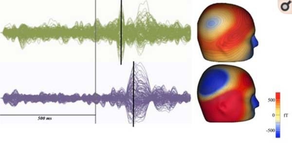 In this photo, two children were given an auditory stimulus to measure their M100 response. The typical child, seen on the top, showed an earlier brain response than the child with ASD. This illustrates that children with ASD generally have delayed auditory processing responses compared to typical children.
EEG - Figure 2
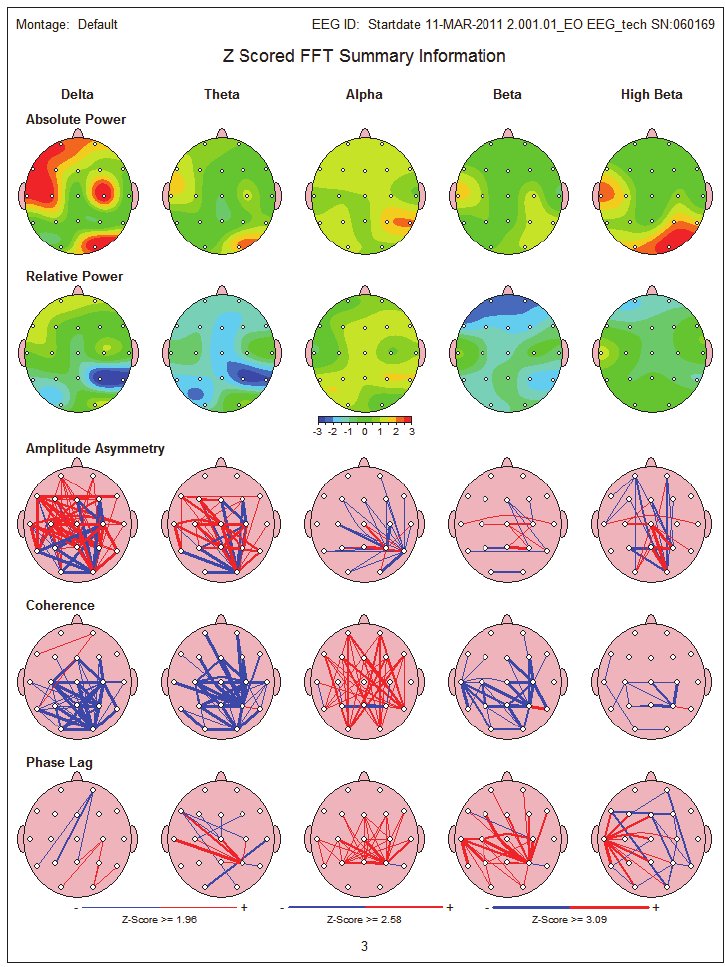 This photo clearly displays how a typical child’s brain activity and energy waves are more evenly spread across the brain, while the ASD child has their brain activity focused in one area and is unevenly spread through the brain.
EEG - Figure 3
This photo clearly displays how a typical child’s brain activity and energy waves are more evenly spread across the brain, while the ASD child has their brain activity focused in one area and is unevenly spread through the brain.
EEG - Figure 3
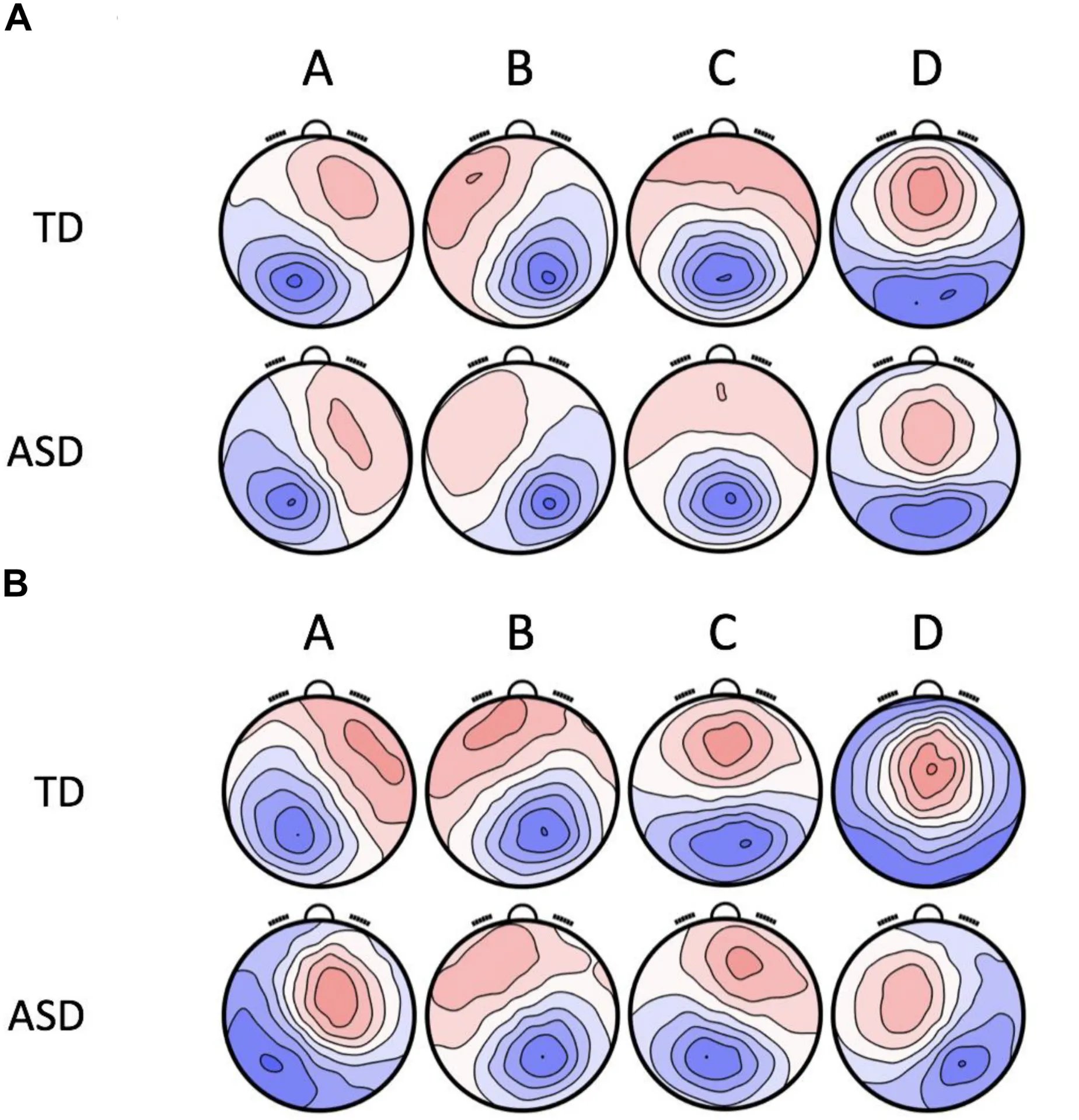 This photo shows topographies of microstates in the brain of typically developing (TD) children and children with ASD. Microstates are patterns of brain activity across the scalp that are brief and stable. They usually last around 80-120 milliseconds before turning into a different pattern. They are used to observe how the brain organizes information at a large-scale network level. In the photo above, the red represents positive voltage and the blue represents negative voltage. If you closely observe the microstates of the children with ASD, you can see that they have shifted peaks, and have less symmetry than the microstates of TD children. Since microstates reflect how the brain connects, organizes, and processes its information, the ASD diagram shows how they organize and process information differently.
Conclusion
Conclusively, MEGs and EEGs can analyze the brain of an individual with ASD to assess their emotions and provide helpful neurofeedback. This neurofeedback can aid the individual to receive a better quality of life.
Citations
- McClure\, Connor. “Unveiling Hidden Struggles: Top 10 Autism Challenges Explored - My Autism Mind.” Myautismmind.com, 18 Aug. 2023, myautismmind.com/autism-challenges/.
- “Neurofeedback.” Psychology Today, 2017, www.psychologytoday.com/us/therapy-types/neurofeedback?msockid=14b744f923c36abb09a651dd22776b8b. Accessed 4 Mar. 2026.
- MindBrain. “Neurofeedback as a Treatment for Autism Spectrum Disorder: A Psychiatrist’s Perspective - 2025.” MindBrain - Mental Health Clinic, 18 Feb. 2025, mindbraintms.com/neurofeedback-autism-treatment-2025/. Accessed 4 Mar. 2026.
- Lin\, Hsiang-Yuan\, and Meng-Chuan Lai. “The Neuroradiology of Autism: Framing Neuroimaging Investigations of the Autistic Brain Based on the US NIMH Research Domain Criteria.” Neurodevelopmental Pediatrics, 2023, pp. 269–282, https://doi.org/10.1007/978-3-031-20792-1_16. Accessed 4 Mar. 2026.
- Adams\, Matthew. “News-Medical.” News-Medical, 27 Nov. 2024, www.news-medical.net/health/Electroencephalogram-%28EEG%29-Systems-Explained.aspx.
- “MEG vs. EEG: What’s the Difference in Brain Measurement?” Biology Insights, 31 July 2025, biologyinsights.com/meg-vs-eeg-whats-the-difference-in-brain-measurement/.
- “What Is MEG?” Ki.se, 2024, ki.se/en/research/research-infrastructure-and-environments/core-facilities-for-research/natmeg-core-facility/what-is-meg.
- Cleveland Clinic. “Magnetoencephalography (MEG).” Cleveland Clinic, 30 Mar. 2023, my.clevelandclinic.org/health/diagnostics/17218-magnetoencephalography-meg.
- “Monarch House Calgary - Monarch House | Canadian Autism Experts | Services & Therapy.” Monarch House | Canadian Autism Experts | Services & Therapy - You Belong, Nov. 2024, monarchhouse.ca/calgary/. Accessed 4 Mar. 2026.
- Parent’s Guide to Electroencephalograms (EEG) for Children with Autism These Materials Are the Product of On-Going Activities of the Autism Speaks Autism.
-“Autistic Brain Scan.” Animalia-Life.club, 2026, animalia-life.club/qa/pictures/autistic-brain-scan. Accessed 4 Mar. 2026.
- Frontiers. “Frontiers | Peer Reviewed Articles - Open Access Journals.” Frontiers, 2024, www.frontiersin.org/.
- Port\, Russell G\, et al. “Prospective MEG Biomarkers in ASD: Pre-Clinical Evidence and Clinical Promise of Electrophysiological Signatures.” The Yale Journal of Biology and Medicine, vol. 88, no. 1, 4 Mar. 2015, p. 25, pmc.ncbi.nlm.nih.gov/articles/PMC4345535/. Accessed 4 Mar. 2026.
- ResearchGate. “ResearchGate | Share and Discover Research.” ResearchGate, ResearchGate, 2024, www.researchgate.net/.
Acknowledgement
I extend my gratitude to my coordinator, Mr. Maruyama. I also would like to thank my parents for supporting me throughout this project. I am grateful to all those who made my project possible.