Using CRISPR and artificial environments to alter stem cells to create organs
Grade 6
Presentation
No video provided
Problem
What is the problem?
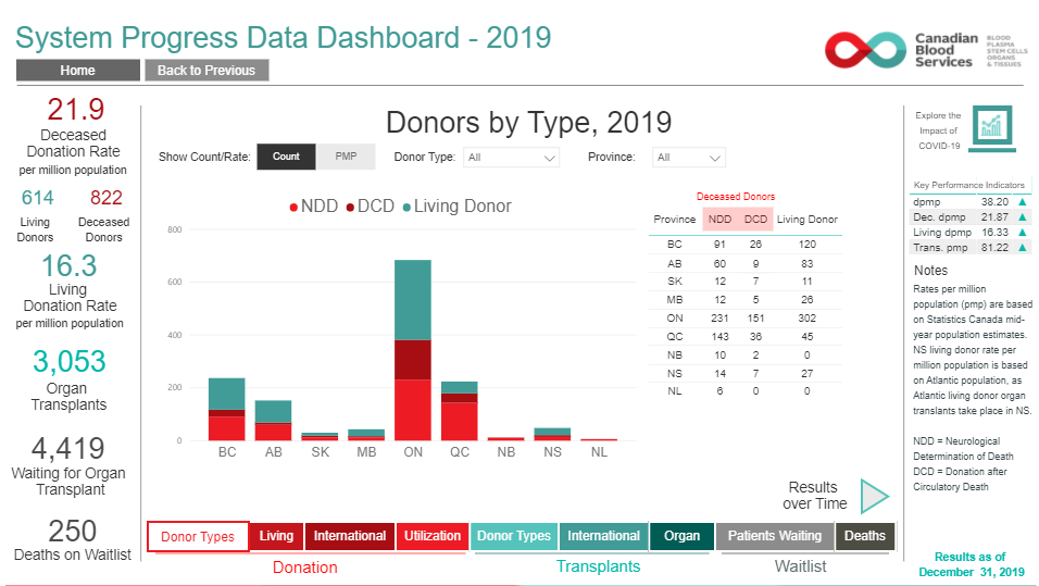
Many people have disfunctional organs and they require an organ from a donor. If you look at the chart below you see that many provinces don't have the organs neccesary for treatment. This problem increases mortality rates by 42% because there are no willing people that will donate the neccesary organs required for treatment meaning that horrible decisions will have to be made for who gets to live from the low amount of organs available. With this new way to create organs we will be able to ensure that everyone waiting for an organs will be able to get the treatment needed for their survival. In 2019 alone 250 people died in Alberta just because there are not enough donors. Although some people strive to help pursuade people to become donors we will need a backup plan just in case there are not enough donors in the future.
Why don't the countries that have lots of organs give some to countries that don't
That reason is because organs aren't so easy to transport to different countries. Organs can't exactly be well stored for later use, but one wasy to transport it is by using cryogenics. Cryogenics is a wasy to preserve organs and bodies until they can be revived or use later. But this method of transportation requires lots of money and time. Something that we don't have a lot of when it comes to patients that require organs quickly. Which is why the method of creating our own organs in hospitals will be a much more efficient source rather than waiting for possibly weeks without a donor. While many countries use this method of supplying countries it is not recommended. Once this is approved of medical agencies we will have more than enough organs needed to help waiting patients.
Is gene editing (CRISPR) even allowed in Canada
The only place that you can currrently access gene editing is in hospitals, this is because that right now CRISPR is being used to help treat sickle cell disease in patients. Although regular civilians have no access to the exponential powers and capabilities of CRISPR or gene editing maybe in the future it will be allowed for researching. If used unresponsibly the powers of CRISPR could have immense negative effects on society which could lead to the demise of our race or create an unstoppable that will wipe out all living things on Earth. Which is why almost all countries forbid the use of gene editing and could lead to major consequenses such as 10 year prison sentence that has been made assured by the Assisted Human Reproduction act. But don't worry, CRISPR and gene editing is heavily guarded to ensure that there is no theft.
Won't the body reject the organ?
Usually if you need a donated organ you will have to get it from someone who is the same blood type of you. This is because your body was made to be adapted to a certain blood type because every single blood type has some unique abliities or is better at some things than other blood types. Without that exact blood type your body would not be functional and could lead to things such as death. Our immune system has adapted and is used to protecting our blood type, so transfusing the normal blood type with an unknown one to your immune system could obviously cause major damage to the body. An example to match the devastating effects of wrong blood transfutions is a malfunctioning nuke that has instead gone to attack the place in which it originated from which you could imagine would not be good for any biotic thing in the surrounding. The immune system then attacks the body because of the unknown blood type with nothing that the body can use to protect itself, this can lead to major consequencel such as death. However some blood types have incredibly unique abilities, the blood type AB+ (positive) is known as the universal receiver. This is because it is able to receive any blood type with a very little chance of things potentially going wrong. While blood type O- is known as the universal donor. This is because the blood type O- has no antigens which is what your immune system uses to know which blood type is safe and which is unknown. You might be thinking, won't the immune system get suspicious that there are no antigens to identify itself. It won't becuase the heart is constantly pumping blood in it's four chambers which is always mixing blood through arteries and veins mixing it with the normal antigen. This problem can be fixed by sparing a few stem cells and put them into a nutrient rich fluid that mimics the environment of blood naturally changing it's form into a blood cell a type of speacialized cell. This creates the purest type of blood that is not yet any blood type that can be donated to anyone.
Method
How can it be done?
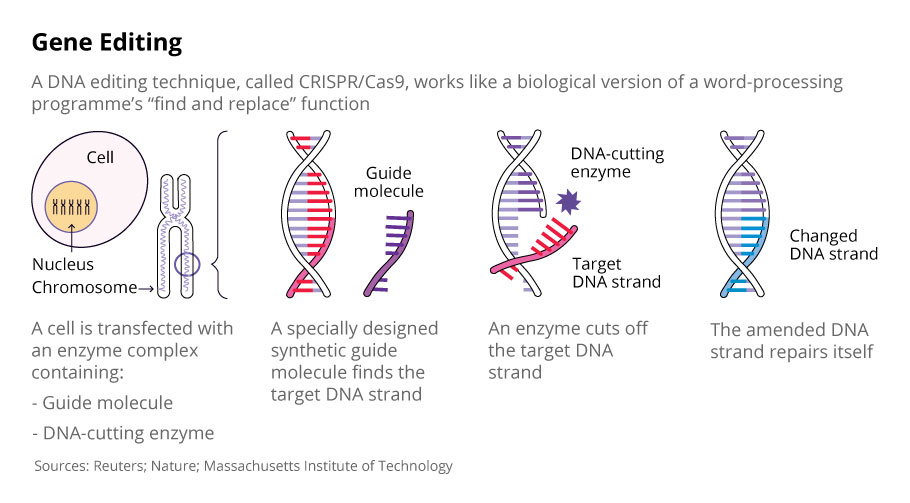
First we can farm our own stem cells or have a willing donor donate stem cells instead of taking whole organs. Then we will have two choices, to use genetic engineering (CRISPR) or we will use artifical environments to change the state of the stem cell to turn it into the cell that is needed to build the organ. So basically if you use genetic engineering you would use a special protein called Cas-9. Cas-9 is a protein that can be found in many bacteria, what it does is constantly check the bacteria's DNA (Deoxyriboucleic acid) to make sure that it hasen't been infected by an organism such as a bacteriophage. Let's say that it did, what it would then do is cut only that piece of infected DNA (Deoxyribonucleic acid) and replace it with a healthy strand of DNA. This exact method will be applied to changing stem cells. If a stem cell is about to change naturally it would change a piece of DNA and changes it so that it slowly changes into a cell such as a tissue cell or a epithelial cell (The lining cell of the lungs and many other organs). Once we have enough stem cells we will be able to create the organ such as a liver or a heart before the previous organ fails.
The process:
First someone has to donate stem cells or we can create our own
Naturally:
- Then if we choose to manipulate the stem cells naturally we would have to make artificial environments
- To create an environment we will have to simulate a similar environment of which the cell would be in if it were to become some type of speacialized cell
- But when it is a stem cell it is in it's DNA to keep on replicating itself so that it can create an organ
- Eventually it would become used to these environment changing itself or creating a daughter cell that specializes in something different by becoming a specialized cell
Using Genetic Modification:
- If we choose to use genetic modification then we will have to aquire a Cas-9 Protein
- Then we will have to study what makes a stem cell change into the specialized cell that we want
- Once we find the gene that makes a specialized cell such as a brain cell the gene that makes it what it is we will replicate it using a synthetic biological circuit or the CRISPR
- (A synthetic biological circuit is an application of synthetic biology in which the biological parts in a cell are being used to and designed to perform logical functions)
- While we wait to study the gene the stem cell will naturally replicate itself creating many more stem cells and daughter cells
- Once we find the specific gene we will replicate it and put them in Cas-9 proteins
- What will then happen is that we will also take out the gene that keeps a stem cell what it is so that it can cut that part out to replace it with the gene that will turn it into a specialized cell
How does Cas-9 work?
Lets say that a bacteriophage attacks a bacteria (bacteriophage is a type of virus that attacks bacteria), then it injects it's own DNA into the bacteria. The DNA injected into the bacteria then creates a viroplasm which is basically a virus factory that uses the bacteria's resources to replicate the bacteriophage. This is when Cas-9 comes in, Cas-9's job in the bacteria is to constantly check the bacteria's DNA to ensure that there is no tamperment in it's DNA. Cas-9 acts like a pair of genetic scissors because once it finds a piece of DNA that it does not recognize it will then very carefully cut that exact piece out and it starts to recreate the piece of DNA lost and it then replaces the infected gene with the new replicated gene. If the Cas-9 protein did not do this then the bacteriophage would have kept on replacating and releasing an enzyme that puches holes in the bacteria to free the bacteriophages and repeat the process all over again. This process will similarily be applied to create organs.
What is next?
Because Cas-9 and CRISPR technologies are only available to hospitals we would need approval of many doctors and specialists before it can actually be used to create organs. But even if it takes a few years for this method of organ creation it would all be worth it in the end because it will save many people. Perhaps this method will in the future be used for other technologies such as suring sickle cells or cancer.
Research
Introduction
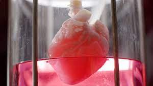
Between 2003 and 2012 organs donors heve grew every year in Canada. This kept happening until 2011 when organ donors numbers started to decease, ever since then there has been fewer and fewer donors and that means more and more paitients requiring treatment just awaiting for a donor to donate an organ. Many years people have tried to adveritise the dangerous levels of organ donors and still not many people help the cause. That is why I thought about organ replication using genetic engineering and modification. This could save the lives of millions of peopple around the globe, and genetic modification can also be used for the lengthening of human life to the accepted age of 120. All of the organs tht are not currently needed can be kept safe in a cryogenics chamber that will keep them preserved until the organ is required for treatment. Currently there are 2 known ways to create or chage an organ, one being xenotransplantation which is taking an organ from a different species and changing it so that it is usable by the recipient or by 3-D printing an organ.
What is Genetic Engineering
Genetic Engineering (Genetic modification) is a process in a laboratory or hospital based machines or living organisms to alter the genetic code in a living organism. This may require changing the base pairs Adenine-Thymine and or Guanine-Cytosine (A-T or G-C) meaning deleting an area of Deoxyribonucleic acid (DNA) and replacing it with a new segment of Deoxyribonucleic acid (DNA). Usually when genetic engineering or modification is used it cuts out the damaged or averse gene to remove or give the organism a desired trait. This transition of genomes can also involve organism to organism gene transitions by directly taking the gene from the animal or by replicaing the gene by means of DNA replication. DNA replication is when you unzip the double helix and unwound it then the separated strand which is shown here as turquoise acts as a template or a base for replicating a partener gene creating an exact copy hence the name DNA replication. DNA constists of a double helix which includes a complementary strand at the
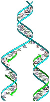
back of the nucleotides (adenine, guanine, cytosine & thymine) called the sugar phosphate backbone that keeps all of the nucleotides in place therefore preventing radiation easily knocking it out of place. This technique can be used to modify stem cells as well, it can be done by identifying what cells are in the organ we need first. Let's say that we are trying to replicace a heart. We know that there are many types of cells with the most abundant probably being a muscle cell. To create a muscle cell we will probably have to place it in an high protein and nutrient environment, with muscles cells being made out of proteins the environment will stimulate the stem cell to evolve into a muscle cell. A segment to the process that I have not yet explained is that we are also able to 3-D print organs using the specialized cells. This might seem a little messed up but organs are just different cells in different arangments and sizes with each of their unique abilities taking part to make an organ functional an usable.
Now if you look at the diagram to the right you will see green arrows spanning the seperated strand. The green arrows represent the places that have been replicated by using different nucleotides. It also represents where the phosphate sugar backbone would go to fully replicate the gene making it usable to program cells into what we want it to become. Another form of genetic modificationis by using a protein popular in many bacteria, Cas-9. Cas-9 is unique because it has the ability to cut out a gene perfectly, this is why it is the chosen for genetic engineering (modification). Scientists and speacialists are thinking of applying genetic engineering to help create cancer cures, to cure sickle cells and using it to create healthier fruits and vegtables or genetically modified plants (GMO genetically modified organism) and livestock.
What is Cas-9 and how does it work
Cas-9 works like a pair of genetic scissors that is programmed to only cut out the infected or damaged piece. to explain how this works let's say that a bacteriophage (phage) attacks a bacterium. It immeadiately starts to inject it's own Deoxyribonucleic acid (DNA), after the injection the bacteriophage leaves the bacterium to repeat the process. When this happens the bacterium is now being forced to produce more of the bacteriophages. What then happens is that after many bacteriophages are produced they start to secrete a special enzyme called lytic enzymes or lysins. Lytic enzymes are highly evoled molecules or enzymes that is produced by bacteriophages or phages that disintegrate the bacterial cell wall allowing the bacteriophages release to repeat the process all over again. This process of destroying bacteria is so effective that 40% of bacteria in the ocean die each day, bacteria in the ocean are still existing because they have evolved to reproduce incredibly quick producing about another copy of itself every 30 minutes. The reason bacterium's aren't able to protect themselves is because their defence systems are too weak against the abilities of the bacteriophage. But sometimes the bacterium is able to react in time to repel the bacteriophage attack, it does this by blocking the DNA entry. How it blocks DNA entry is by adding a simple attachment to a suitable surface receptor such as an MHC class 2 window (a basic molecule that displays what a cell has produced). Superinfection exclusion (Sie) system can help block bacteriophage DNA by membrane anchored or membrane accosiated protein is installed to the bacterium. But let's say that a bacteriophage get's past the bacterium's defences and the Sie system so that it is able to inject it's DNA, before it injects the complete DNA for the bacterium to produce let's say that a protein from the Sie system manages to deflect the bacteriophage.
Then the bacterium would be safe because the complete DNA was not yet injected, after the attack the bacterium would store the bacteriophages DNA into it's DNA archive until it is required to protect itself from a bacteriophage attack. Let's say that a bacteriophage did attack the bacterium again then it would activate the Sie system and it's other defences but if it still manages to hijack the cell then the bacterium would create a RNA copy of the bacteriophages DNA from the previous attack and load the RNA into an incredibly powerful protein called Cas-9. What Cas-9 does is search the bacterium's DNA to check if there is any of the bacteriophage DNA that is forcing the bacterium to produce more bacteriophages. If it does come across a piece of the bacteriophage's DNA it will then cut the piece like a pair of genetic scissors and replace it with a healthy piece of DNA. But wait, how will the Cas-9 protein know when it comes across a piece of bacteriophage DNA? Remeber earlier when the bacterium produced the RNA copy of the bacteriophages DNA from the DNA archives. It compares that RNA piece calld the guide molecule to the DNA of the bacterium, finding the exact piece and cutting it out to then replace it with the bacteriums own DNA. After this the Cas-9 protein is done it's job and is stored for later requirment, the bacteriophage attack has been prevented and the bacterium is now safe.
How Can Genetic Engineering Be Applied to Organ Creation
Genetic engineering can be used to create organs by taking stem cells or organs from other animals and modifying them. If we are to take an organ from an animal we will have to make changes depending on which animal we take it from, currently scientists say that the best organ to harvest from are pigs. This method of organ transplantation is called xenotransplantation which is when organs, tissues and blood is harvested from a different species. There are many problems with harvesting organs from other animals such as immune defences size and molecular incompatibility and much more, all of these problems lead to xenograft rejection. But through the advancments in genetic engineering it is now possible to prevent all problems of incompatibility between the domesic pig and a human. Currently there are several techniques to safetly transplant the organ of a pig to a person each of the following will be listed: pronuclear and cytoplasmic microinjection , somatic cell nuclear transfer (SCNT) and viral transduction of DNA. The techniques listed above help prevent xenograft rejection and safely transplants the organ from the domestic pig to a human. The Sleeping Beauty microinjection technique is a germline transgenesis (transferece of a DNA segment to give the gene to an offspring) but instead it will be used to inject the gene into stem cells to create the desired organ. Another reliable source is the piggyBac microinjection for easy and efficent genomic insertion. The piggyBac system uses the exogenous DNA (DNA originating outside of the organism of study, it is usually partially completed around dead cells) and microinjecting it into embryo's which will automatically multiply when DNA is inserted. Cas-9 is the protein that we will be using to make specific gene modifications to make small ajustments in the genomes. Currently there are 4 known ways in which the body will reject the organ in xenograft rejection: Hyperacute Rejection (HAR), Delayed Xenograft rejection (DXR), Acute Celluar Rejection (ACR) and Chronic Rejection (CR).
Hyperacute Rejection:
Hyperacute rejection occurs within about 3-5 minutes after the transplantation of the new organ meaning that the new organ has failed. The primary role that hyperacute rejection plays is mostly done by endothelial damage and loss of a variety of its biological properties. Endothelial damage happens when the large blood vessels on or close to the hearts surface start to constrict narrowing instead of dilating (opening), this tends to cause chronic chest pain. Inside the organ one thing that commonly happens is intravascular coagulation (the blood in the vessels and arterys starts to thicken, this technique is usually used to make scabs on the surface of the skin), many infiltrations of the bodys immune cells start to begin invading the organ. This invasion is mostly composed of neutrophils (a very vital cell in the immune system that releases acids to kill bacteria), the neutrophils start to attack the transplanted organ leading to necrosis (cell death). One of the main reason's that hyperacute rejection happens is the natural presence of preformed (meaning existing before the transplantation) antibodies inside the human's plasma that recognizes the many differences that were not fixed making the immune system think that the new organ is an enemy.
Antibodies in the human plasma reconize a gal antigen on the surface of endothelial cells, what then happens is that the gal epitope is now starting to be formed by the bonding of the galactose (natural sugar) to N-acetyllactosamine a component of a variety of glycoproteins that works and functions like a carbohydrate anitigen that plays roles in normal celluar recognition as well as in maligant transformation (a process in which cells begin to aquire some properties of cancer) and metastasis when cancer cells move in the body from the place in which it originated in. The bondage of the Gal epitome and the N-acetyllactosamine is bonded by a glycosidic linkage which is a chemical bond that creates a connection between a carbohydrate or sugar molecule to a differnet group such as celluose. The catalyst that will create this reaction is a galactosyltransferase or a GGTA1 enzyme, the binding and recognition of a Gal antigen by xenoreactive antibodies that activate the complement system a system composing of proteins that cripple and destroys bacteria and viruses. The complement system then starts to begin a cascade that activates other proteins to eventually create a membrane attack complex or MAC, the membrane attack complex is basically a membrane penetration by proteins to form a membrane channel which spews out the insides of the bacter or virus ultimately resulting in cell lysis which means the breakdown of a cell due to damage or inffliction to the outer membrane.
To ensure that the tranplanted organ doesn't get rejected by hyperacute rejection is by removing the Gal antigen off of the xenograft cell or cell surfaces to ensure the safety of the recipient. The safest and best method of doing this is by removing the Gal epitopes is by inactivating the gene that encodes the GGTA1 enzyme, this is because it acts like a catalyst for the Gal epitope that creates the reaction. To do this genetic recombination was applied to replace the GGTA1 gene with a different variant that will prevent the production of the GGTA1 enzyme. In 2001 an experiment was held where pigs were heterozygously (to have inherited a part of the biological parent's gene) inactivated GGTA1 were produced, 1 year later piglets with homozygous (to inherit the same genes as the parent) inactivation of the gene. The first xenotransplantation using hearts from GGTA1 inactivated pigs was done in 2005, the recipients were immunesuppressed baboons. the survival period was about 92 days and the longest surviving graft (taking one piece of a living tissue from an organism and giving it to another organism so they grow as one) in the recipient was 179 days in total. A intracytoplasmic microinjection of Clustered Regulary Interspaced Short Palindromic Repeats or the CRISPR system which is the Cas-9 protein obtained the biallelic knockout of the gene that produce GGTA1 in 3 of 6 piglets.
The microinjection avoids the penetration of the pronuclear membrane which would kill the cell, this increases the survival rate of the microinjected embryo. CRISPR is a very powerful tool and it significantly facilitates and shortens significantly the process of each generation pig by using multiple genetic modifications making the xenotransplantation much easier. However xenoreactive anitbodies are one of the main reasons for hyperacute rejection, the complement system a powerful tool plays a pathophysiological (a disordered processes that are accosiated with diesease or injury) role in hyperacute rejection. Some complement proteins have been found to regulate complement activity, the ones that regard the most to xenotransplantation include proteins: CD46, CD55 and CD59. CD46 is a membrane cofacter protein that protects the cell by blocking the formation of the C3 convertase complex which basically activates other complement proteins that start to begin a cascade crippling the bacteria or viruses defences. CD55 is a decay accellarating factor that regulates cell susceptibility to a complement protein attack, the role it plays is inhibiting the formation of a C3 or C5 convertase and speeds up or accelerates the decay of the platform. CD59 is a membrane inhibitor of reactive lysis which prevents the formation of MAC or Membrane Attack Complex which is the final stage of complements cascade. CD59 protects cells protects cells from lysis by binding to complement C8 and C9 blocking C9 polymerization (the process to create polymers) and cell membrane attack.
Delayed Xenograft Rejection:
A xenograft transplant that avoids hyperacute rejection is subjected to delayed xenograft rejection, this takes place several hours to several days after the organ transplantation. Delayed xenograft rejection is quite similar to hyperacute rejection, the difference is that it does not require the participation of the complement system. Instead it involves immunoglobulinss which measures the amount of antibodies there are in the blood. Immunoglobulins develop much slower and is initiated in the arterial lumen (a hollow passageway in which blood flows through) and not in capillaries or aterioles. During delayed xenograft rejection endothelial cells which is the thin layer covering blood vessels and arteries undergo a type 2 activation, a type 2 activation creates proinflammatory genes, and an increased secretion of chemokines (which are basically receptors for cells) and blood platelet activation. Coagulative disorders happens to be the result of molecular differences in the pig and human coagulation systems, the complement protein CD39 is an ectoenzyme which is a catalytic membrane protein around a cell. CD39 is present in the vascular endothelium, a layer of endothelial cells which lays in the inner cellular lining of arteries, veins and capillaries. CD39 plays an important role in coagulative responses and inflammatory processes, it's activity inhibits platelet aggregation which is triggered by adenosine triphosphate (ATP) and adenosine diphosphate (ADP) release. CD39 hydrolyzes ATP and ADP into adenosing monophosphate (AMP), the latter (when a diesease has run it's course and the person is about to die) being a strong platelet aggregation inhibitor which prevents the coagulation system from working killing the person.
Thrombodium or TM is an integral membrane protein on the surface of endothelial cells, it plays a role in coagulative inhibition. TM activates protein C, which is highly anticoagulative, protein C is a protease or a proteolytic enzyme which is a group of enzymes that catalyze the hydolysis of bonds in proteins and polypeptides, this catalyzation is highly effective on peptide bonds. Protein C with protein S as a cofactor (a non protein chemical compound that is required for an enzymes role as a catalyst, inhibits factors Va and VIIIa which are blood coagulation factors are homologous cofactors for prothormbinase (prothormbinase consists of factor Xa which is a serine protease with factor Va a protein cofactor) which are apart of the coagulation system. With factors Va and VIIIa being inhibited the whole enzymatic cascade is inactivated causing clot formation, with the inability of porcine (resembling a pig or pigs) TM able to bind to human thrombin (a special molecule that acts both like a procoagulant and an anticoagulant). With TM not being able to bind to human thrombin prevents protein C activation. In an experiment people have succesfully generated transgenic pigs with hTM gene by using somatic cell nuclear transfer, somatic cell nuclear transfer is when you take a nucleus of a cell with its DNA and put it into a different cell that could be done using microinjection.
It showed tha hTM porcine fibroblasts showed elevated activated protein C production in an in vitro TM coactivity assay. By using GTKO/hCD46/hTM pigs and very high doses of anti CD40 immunosuppressants achieved the longest xenograft survival time of 945 days, in heterotopic (occuring in an abnormal place) pigs to non human primate cardiac xenotransplantation model. The vascular endothelium produces one of the strongest natural extrinsic coagulation pathway inhibitor or TFPI (tissue factor pathway inhibitor), the anticoagulant activity of TFPI composes in reversible inhibition of factor XA and the formation of the Xa/TFPI complex which inhibits the TF (tissue factor)/ VIIa complex. TFPI plays a double role in the process of coagulation inhibiting both the Xa and the TF/ VIIa complex, it is the only inhibitor acting at the early stage of coagulation preventing the formation of thrombin. Porcine TFPI is not a very effective inhibitor of human factor Xa and might be ineffective in deactivating TF.
Cellular Rejection:
Cellular Rejection occurs about within several days after transplantation. The dominant morphological characteristics of such rejection are infiltrations with pre dominant mononuclear cells, located interstitially (of forming or occupying spaces) in the tissues. In the human the celluar response tageting porcene tissues is induced both by Natural Killer cells (NK cells) and by T lymphocytes which are T-cells that help fight cancer and is developed in the bone marrow. Both NK cells and T cells with CD4 and lymphocyes exhibit a very high level of cytotoxicity than CD8 and lymphocytes. The susceptibility and vulnerbility of porcine endothelial cells to lysis are induced by Human NK cells. To the inability of NK cell inhibitory receptors to identify major histocompatibility complex MHC class I molecules which are like windows that let immune cells see what the cell has been producing. The differences between porcine (swine leukocyte antigens) and human (human leukocyte) MHC windows. The introduction of the HLA-E gene (It both inhibits and activates NK cell mediated and also controls cytotoxicity and cytokine production which are like molecules that activate other cells) into porcine endothelial cells has shown to partly protect the porcine endothelial cells in virto. One team of scientists showed that the introduction of the HAL-E gene into pigs not only protects the porcine organs but also against cytotoxicity as well as macrophage (another very vital cell in the immune system that digests and kills bacteria and viruses) cytotoxicity. Another study shows that relative to pig lungs perfused with human blood with the HLA-E gene increased median lung survival rate in vivo and was associated with reduced pulmonary vascular resistance and decreased platelet activation.
Another cause of cytotoxicity leads to lysis of the porcine endothelial cells is the binding of porcine UL-16 binding protein which are realative to MHC class I molecules. UL-16 binding proteins play an important role in activating natural killer cells (NK cell) and plays an important immunomodulary role. Macrophages are a functionally heterogenous (differing in kind) population of mononuclear cells playing an incredibly important role in inflammatory processes as phagocytic cells. They are activated either by xenoreactive (that generates a reaction to material from another species) T cells or by direct interaction between a donor endothelial antigens and macrophage surface receptors. Primate macrophages have been shown to participate in the rejection of some porcine pancreatic islets (a pancreatic cell that produces hormones that are secreted in the blood stream such as insulin and glucagon) with Gal epitopes removed and to phagocytose porcine reb blood cells independently of antibodies or the complement system. Phagocytosis is inhibited by the CD47 surface antigen, protein CD47 is an integrin associated protein and is a member of the Ig (Ig stands for immunoglobulin) superfamily (an immunoglobulin superfamily is a large proterin family of cell surface and soluble proteins) in all tissues. The signal regulatory protein SIRP is present on cell membranes is an inhibiting receptor that is able to recognize CD47. The CD47 and SIRP interaction delivers a phagocytosis inhibiting signal to macrophages. An experiment (jung et al) was taken and it demonstrated that CD47 and SIRP interaction, TFPI expressed by transgenic porcine cells enhanced the CD47 SIRP axis and could possibly improve the CD47 and SIRP signaling and might help overcome macrophages mediated immune rejection.
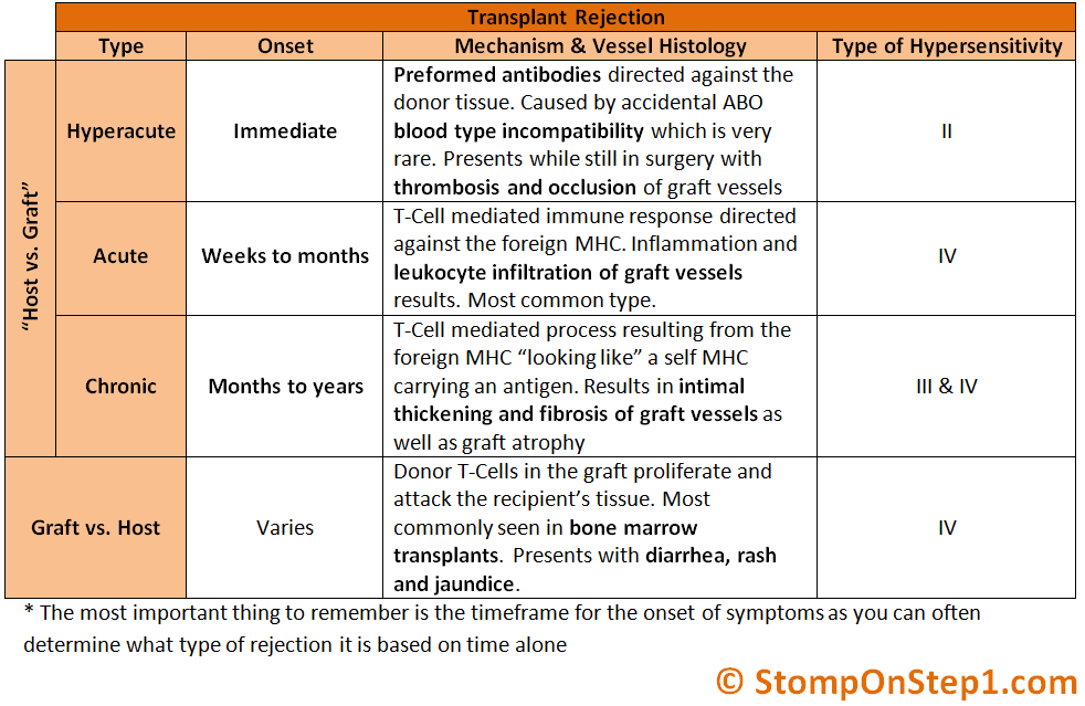
Chronic Rejection:
Chronic Rejection can occur within months or even years after the transplantation, and it is clinically manisfested by progressing failure of a grafted organ or tissue. It is estimated that chronic rejection occurs when additive effects of various harmful factors, very little is known about chronic xenotransplant rejection but it is suggested that one of the intiating factors may be specific antibodies. Scientists also think that an intiative factor may be connected with damage of vascular endothelium by cytotoxic T lymphocytes and specific antibodies. But the intensity of the response is to low than in the case of hyperacute rejection, in cell membranes we see the increased expression of cell adhesion molecules and the tissue factor that activates the coagulation system. Gradual proliferation of the aortic media takes place leading as a consequence to vascular obliteration and organ failure.
How can organs be 3-D printed?
 Printing organs is called bioprinting, this is because instead of using plastic the speacialized printer is proggramed to create an environment in which an organ will be preserved in while in the process of creation. This is a very advanced and complex technique that has been improving over the past few years. 3D printing has lead a very big footprint in medical sciences because it could be used for testing and research of dieseases on actual tissue. Many other solutions to things such as creating bio materials such biomolecules. Every little detail has an essential part in how the organ will turn out, the needle of the 3D printer itself is less than 1 milllimeter in diameter. Human body's themselves are very regenerative but are restrained my various factors such as growth hormones, different tissue types, the body's resources, physical size and age. If you look at the diagram above you will see that biopolymers are required, biopolymers are substances created by living or biotic organisms. Like many polymers biopolymers are made by molecules called monomeric units, monomeric units are just chemicals created by molecules that bind together to form a repeated or unique pattern that becoomes a biopolymer. A bioink which is shown in the diagram above is a combination of biopolymers and cells that has been chosen for organ creation because of it's excellent biocompatibility. The bioink mostly is composed of cell but the biopolymers are added to make the body think that it is real tissue. In the diagram there are two arrows sprouting to in virto and in vivo, in virto means testing outside of a living organism. With in vivo meaning having the organ tested inside of a living organism. Below will be the process of how the organ is going to be made:

The process of organ creation and testing for inkjet:
- A biopolymer or synthetic polymer is used and loaded into the 3D printer
- The needle is incredibly thin for high precision
- A piezoelectric or a thermal (the property of certain bodies to be electrically polarized to create an electric field or potential uder the action mechanical stress) actuator would be needed to create an evaported ripple that will let the bioink droplets to flow through rather than having it clogged
- After proggraming the 3-D printer to create a certain organ it will take time for it to be completed
- To guarentee that a organ will work you can use the methods of in vitro meaning that you test the organ functions outside of a living organism or in vivo meaning that you test the organ inside of a living organism
The process of organ creation for extrusion method:
- For the extrusion method the bioink is loaded into the 3D printer for the needle to extrude
- A pneumatic or a screw would be use to slowly extrude the bioink droplets in a certain order to create an organ
- Mainly high viscosity bioinks are used in extrusion bioprinting
The process of organ creation for laser assisted method:
- A few layers would be needed to protect the bioink and keep maximum precision
- An energy absorbing layer (donor layer) would be needed to absorb the impact of the laser
- A seperate bioink layer will be put under the donor layer or energy absorbing layer
- The donor layer only protects the impact and not the heat so the bioink layer slowly bubbles release the bioink but still keeping ultimate precision
- This technique permits high resolution deposition of where the bioink droplets will be in solid or liquid phases
- this photo polymerzation method can modulate laser assisted bioprinting of biomaterials as well as a variety of different cells without affecting the cells viability and functionality
- The laser pulse keeps on going until the organ is complete
The process of organ creation for steriolithography method
- Layer by layer a photosensitive polymer layer is applied
- The layers are then solidified by using an ultraviolet light eventually creating a multiplex structure in the end
- The organ format to build off of is made by real organ scans such as Magnetic Resonance Imaging (MRI), Computac axial tomography (CAT scan) and Computed tomography (CT scan)
- For printing different pieces of organs such as alveoli and lungs, this is fixed by using different nozzles to create micro ore nano details vital in the alveoli
The history of 3D Bioprinting
The term 3D printing was created by Charles W Hull, he named a method of organ printing called steriolithography. Steriolithography is when you put a thin layer of material over another layer and project a UV light onto the material to harden it that eventually creates a 3D structure. This method was later tested using biomaterials to form 3D scaffold using cells, this was later used with the latest tissue regeneration 3D technique using celluar biology which acronym is FRESH. Also known as Freeform Reversible Embedding of Suspended Hydrogels which was discovered by Professor Adam Feinberg and his feam form Carnegie Mellon Universtiy. This same technique was used to make much more complex biological structures some such as lungs, hearts and soft biomaterials held in a gelatin bag to overcome the constrainsts of soft bioinks. This is one of the most complex processes against the three other techniques which are inkjet, laser-assisted and extrusion. Charles W. Hull.
Using Artificial Environments
 This may not be very understandable because it hasn't actually been tested and was just an idea that appeared when thinking of organ creation. Using Artificial environments to create organs has both it's pros and cons, one pro is that it could facilitate more safety in organ creation, a con is that it could completely backfire and that the organ are dysfunctional. How this works is that stem cells are going to be placed in an environment that will synthesize and simulate the environment that the desired cell would be in and experience. After a while the stem cells will evolve into a speacialized cell. Some environments such as a high nutrient environment might cause the stem cells to turn into a red blood cells, and high protein environments could create muscle cells. The easiest organ to create is a heart because it is mostly composed of muscle cells and there are barely any chemical reactions. To create a heart the stem cells will have to be shaped to create the 4 chamber and all of the veins and arteries to perfectly align with the recipients body. The heart can be shaped by using a Da Vinci surgical system, which is an incredibly precise machine that is used for intricate and complex surgeries. With the heart shaped it can now be put into the solution where the stem cells will gradually change and evolve into the specialized muscle cell. With the heart completely finished it can now be used but first for the heart to work it will have to be hooked up to the neurological system so that the brain is able to control the heart and make it beat and spread blood through out the body. A pro is that this would be quite efficient and would be cheaper than 3-D bioprinting and genetic engineering along with xenotranplantation.
Data
N/A
Conclusion
Conclusion
Although some of these organ creation methods aren't currently being used right now they are being tested so that maybe in the near future it would be accesible by people who require these organs. This presentation was just to show how much the world needs more organs and some methods to create them as well as a new method of Artificial Environments. All of these methods have their pros and cons, such as high cost and incredible precision. But to summarize everything in this presentation organs can be created by using 2 known and tested methods, these are xenotransplantation and 3-D bioprinting. Xenotransplantation has 4 main problems in which the body will reject the xenograft organ: Hyperacute Rejection, Cellular Rejection, Delayed Xenograft Rejection and finally Chronic Rejection. Where as 3-D bioprinting also has some problems such as it can only print low density bioink or that the process is not precise. But all of these problems can be fixed and people in the world will receive the help and support that they need.
Citations
Bioartificial Organ Manufacturing Technologies - PMC

National Institutes of Health (NIH) (.gov)
https://www.ncbi.nlm.nih.gov › articles › PMC6322143
https://www.ncbi.nlm.nih.gov/pmc/articles/PMC9088731/
https://www.ncbi.nlm.nih.gov/pmc/articles/PMC3510378/
https://www.ncbi.nlm.nih.gov/pmc/articles/PMC4982070/

https://en.wikipedia.org › wiki › N-Acetyllactosamine
hyperacute rejection - Google Search
The UL16-binding proteins, a novel family of MHC class I ...

National Institutes of Health (NIH) (.gov)
https://pubmed.ncbi.nlm.nih.gov › ...
HLA-E major histocompatibility complex, class I, E [ (human)]
National Institutes of Health (NIH) (.gov)
https://www.ncbi.nlm.nih.gov › gene
Cloning animals by somatic cell nuclear transfer - NCBI
National Institutes of Health (NIH) (.gov)
https://www.ncbi.nlm.nih.gov › articles › PMC521203
Prothrombinase - Wikipedia
https://en.wikipedia.org › wiki › Prothrombinase
Acknowledgement
Acknowledgements
All of the people that are listed below have helped me so much to support and help me while I have don science fair and I want to thank all of them for helping me through it all:
My Dad Zalaan My Grandma
My Mom My Uncle My Sister
Mrs Thomas Tayson My Cousins
Mrs Strohbach My Grandpa Claire