How can we use technology to predict and help treat Myocardial Ischemia disease?
Grade 7
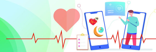
Presentation
No video provided
Problem
Ischemia is a major issue these days. The most severe forms are cerebral (affecting the brain), myocardial (affecting the heart), mesenteric (affecting the intestines), limb, and renal (affecting the kidneys). Myocardial ischemia, which happens when blood supply to the heart muscle is reduced by a partial or total blockage of the heart's arteries, is one of the most fatal types. Numerous issues can arise from myocardial ischemia, and the only ways to cure it are with medication, surgery, or lifestyle modifications. My question is, now that technology is evolving and that there are more fields out there which use new different types of technology to predict Myocardial ischemia disease, using the factors on how the machines predict if someone has the disease, can we analyze the data, compare it to the symptoms of ischemia diseases, compare all technology with symptoms, in order to come up with a way to increase the knowledge about treatment?
During this research project I will research on how ischemia disease forms, what are the risk factors, symptoms, how it affects one’s body, treatments already active now and how it affects one’s daily lifestyle. Then using that research collected, I will research about different types of technology, what they specialize in, how they detect if someone has Myocardial ischemia disease, how the technology works, how they compare to the symptoms and then compare all of technology and symptoms in similar and different aspects. I will compare the outcomes, look for a theme among them all, and use charts and diagrams to formulate a recommendation for more care.
Method
Background research: Ischemia disease occurs when there is a restriction in the blood flow to any organ, muscle group, or tissue in the body. This results in a deficiency of oxygen, which is necessary for the proper functioning of cellular metabolism. Depending on where the blood flow is restricted, there are many forms of ischemia disease. This can occur in any part of the body, including the legs, the heart, and the brain. While certain forms of ischemia are more deadly than others, all forms carry a significant danger of some kind if not treated appropriately. These conditions can result in strokes and heart attacks, among other potentially fatal conditions. Ischemia disease can have a significant impact on day-to-day living. The only options for treatment are surgery, medication, or lifestyle modifications. Treatment is very individualized and varies depending on the type of ischemia, how serious it is, and the individual's typical daily activities.
Technology nowadays is growing; many new types of technology are being implemented in the medical field.
An electrocardiogram (ECG) is a test that records the electrical activity of the heart, it shows abnormal things such as changes in heart, signs of ischemia, or previous myocardial infarction. It is widely used to test if one has ischemia disease.
Echocardiography uses ultrasound waves to create images of the heart chamber, surrounding structures, and valves. It accesses the heart pumping action, and it can predict if one has Myocardial ischemia by observing the area that is not getting enough blood.
Cardiac MRI (Magnetic Resonance Imaging) provides detailed images of the heart's structure and function. It is more specifically used for examining and accessing the heart muscle after a heart attack, along with detecting reduced blood flow.
What are the treatments provided for people with Myocardial Ischemia? There is no specific treatment for Myocardial ischemia, but doctors say the best things to do are to change daily lifestyle, take specific medications, or get surgery.
Since technology is evolving using the symptoms there are for ischemia disease and how each piece of technology checks to see if the patient has Ischemia disease, after gathering research on each piece of technology, comparing them on a chart with the symptoms and comparing all pieces of technology with the symptoms, can I find a way to enhance the treatment for ischemia disease?
Research
ISCHEMIA DISEASE
What is Ischemia Disease?
Ischemia is a medical disorder where a portion of the body has a decrease in blood flow due to a blockage or constriction of the arteries supplying that area. The tissue or organ in question may become damaged or cease to function as a result of the reduced blood flow, which also results in a shortage of nutrients and oxygen. The shortage of oxygen will also affect the cellular metabolism of the patient. The type of ischemia the patient has depends on where the clot of blood is, it can happen almost anywhere in the body, but is most found in the heart, legs, brain, intestines, limbs, and kidneys. If a patient has ischemia, their lifestyle will change drastically as the ischemia affects the patient’s body in many ways.
What are the symptoms?
The symptoms of having Myocardial ischemia disease are:
Having neck, jaw, shoulder, arm or ear pain; It has been newly discovered that having neck, jaw or pain can mean early signs or heart problems prior to a heart attack. It is not fully understood well but it is said that symptoms of having myocardial ischemia disease can be having neck, jaw, shoulder, arm or ear pain because of the phenomenon called referred pain. This occurs when pain originating of the body is felt in another part. In the hearts case, it does not have a direct way to signal distress through pain because of less pain receptors, compared to other areas, so when there is a problem such as reduced blood flow, the body may interpret it in different areas of the body. The body parts mentioned above are all supplied by nerved from the same general region of the spinal cord (the cervical and upper thoracic spinal segments). When the heart is going through Myocardial Ischemia, it sends signals to these areas. However, the brain may have difficulty pinpointing the exact origin of the pain, so instead it perceives that the pain is coming from the areas of the body that are innervated by the same spinal levels as the heart. That is why pain in the neck, jaw, shoulder, arm or ear may indicate someone has myocardial ischemia disease.
Having a fast heartbeat or angina; If the coronary arteries are seriously block because of having myocardial ischemia, blood flow may not be adequate for any increase demand, stuff like exercise or being emotionally upset. If the heart muscle can’t get enough oxygen (myocardial ischemia) symptoms such as shortness of breath and angina (chest pain). A fast heart rate can also mean myocardial ischemia because of electrical instability, which is ischemia disrupting the normal electric pathways to the heart, which could potentially lead to arrhythmias, including those that cause of a heartbeat. It can also be a stress response, which is Myocardial ischemia triggering a stress response in the body releasing adrenaline and other stress hormones. These stress hormones then prepare for “fight” or “flight” by increasing the heart rate. Angina is a direct symptom of myocardial ischemia; it means the heart muscle isn’t receiving enough oxygen-rich blood. This discrepancy between oxygen supply and demand causes a type of chest pain of discomfort know as angina pectoris.
Having a shortness of breath when physically active; This could mean one has myocardial Ischemia because it could mean since they might have myocardial ischemia, the heart has a reduced capacity to meet the increased oxygen demands of the body during and exercise. When someone is engaged in exercise, the muscle requires more oxygen to produce the energy needed for movement. The increased demand for oxygen forces a corresponding increase in the amount of blood the heart pumps per minute and the respiratory rate to supply the muscle with oxygen-rich blood and to remove carbon dioxide.
Having Nausea and vomiting; If someone has nausea or are vomiting, it can mean they have myocardia, Ischemia disease because it can mean that the ischemia is triggering a response from the autonomic nervous system, which controls involuntary bodily functions including digestion. Activation from this could lead to nausea. The vomiting could be caused from a pain response, which is the sever pain from the ischemia triggering a nausea response. The body’s reaction to the intense pain, especially if its not localized, can lead to a feeling of sickness or an urge to vomit.
Having Fatigue; Due to the less nutrients and oxygen reaching the heart muscle because the myocardial ischemia disease, since it is crucial for it to function effectively, it can lead to a feeling of overall fatigue, because the body’s tissues are not receiving enough oxygenated blood to meet their metabolic needs, especially during physical activity. In response to the reduced blood flow, the heart may have to work harder to supply the body with the necessary blood. The increased effort can deplete the heart of energy more quickly, leading to a sense of fatigue. There are many other reasons someone may experience fatigue, but those are the most related ones to myocardial ischemia disease.
While most people who have myocardial ischemia disease deal with symptoms, there are some people who don’t have symptoms for the disease, but still have it. This is called silent Ischemia and is most found in diabetes patients, but anyone could have it. The term "silent ischemia" describes a condition known as myocardial ischemia, which is characterized by a decrease in blood supply to the heart muscle without the usual signs of angina, such as pain or discomfort in the chest, Due to the illness being typically remains undiagnosed and untreated because of its lack of obvious symptoms, there is a higher chance of developing more severe cardiac issues, such as a heart attack or sudden cardiac death. So, it is very important to get it checked, before it is too late!
What is specifically Myocardial ischemia disease?
When there is insufficient blood flow to the heart, the heart muscle is deprived of oxygen, leading to myocardial ischemia. The cause of the decreased blood flow is typically a partial or total blockage of the coronary arteries in your heart.
Myocardial ischemia happens when a deposit of plaque (atherosclerosis) partially or completely blocks a coronary artery, preventing blood flow to the heart muscle (myocardium). You may get a myocardial infarction, or heart attack, if the plaques burst.
The condition known as cardiac ischemia or myocardial ischemia decreases the heart muscle's capacity to pump blood. An acute, significant blockage of an artery in the heart may cause a heart attack. In addition, myocardial ischemia may result in dangerously irregular cardiac beats.
How does Myocardial ischemia disease affect the body?
Myocardial Ischemia affects the body by making it harder to function properly daily. The effects of myocardial ischemia on the body can highly depend and vary based on the severity. Some of the key impacts include, Angina, shortness of breath, Arrhythmias, Heart attack (myocardial infarction), reduced exercise tolerance, Heart failure, and an increased risk of sudden cardiac death.
Exercise becomes challenging for those with myocardial ischemia, particularly in the winter. You may have myocardial ischemia symptoms with decreasing activity as your situation worsens. Going up a flight of steps might become challenging over time. At some point, you could even have symptoms while you're sleeping.
The effects of myocardial ischemia on the body highlight how crucial it is to identify, treat, and prevent coronary artery disease as soon as possible. Medications to increase blood flow and decrease clotting, lifestyle modifications, and operations like bypass surgery or angioplasty are typical methods to treat ischemia and lessen its negative effects on the body.
How does cellular metabolism get affected by Myocardial Ischemia disease?
What is Cellular metabolism?
The intricate series of chemical processes that are necessary for life to exist within cells is referred to as cellular metabolism. For cells to grow, maintain their structural stability, and react to their surroundings, these responses are essential. Nutrients are converted into energy and cellular components during cellular metabolism, and metabolic wastes are removed. It includes both the creation of every molecule required by the cells (anabolism) and the breakdown of molecules to provide energy (catabolism).
How does Cellular metabolism work?
All of the chemical processes that occur inside of a cell to produce energy, make essential molecules, and control waste are together referred to as cellular metabolism. A summary of the steps are:
Breaking Down Nutrients for Energy: A process known as catabolism occurs in cells when they take in nutrients like carbohydrates and fats. As a result, ATP (adenosine triphosphate), which the cell requires for a variety of processes, is produced.
Glycolysis and Cellular Respiration: The first step in the breakdown process is glycolysis, in which the sugar glucose is broken down into pyruvate and a tiny quantity of ATP. Pyruvate reaches the mitochondria for additional processing (aerobic respiration) if oxygen is present, which results in a significant amount of ATP synthesis. In the absence of oxygen, cells turn to fermentation, which is less effective in generating ATP.
Building Up Cellular Components (Anabolism): In order to construct larger, more complex molecules like proteins, nucleic acids, and lipids, cells also employ part of the energy and smaller molecules created during catabolism. The anabolic process is essential for the growth, multiplication, and repair of cells.
Regulation and Balance: To guarantee that the proper quantity of energy and materials are accessible in accordance with the needs of the cell, the entire metabolic process is strictly regulated. To keep the cell healthy and functioning, this entails regulating the activity of enzymes and striking a balance between energy production and consumption.
Cellular metabolism can be compared to a delicate balancing act in which cells utilize the energy they obtain from the breakdown of nutrients to produce energy and then use some of the breakdown products to assemble the necessary components, all the while carefully regulating the process to satisfy their current and future needs.
How does myocardial ischemia affect cellular metabolism?
Reduced blood flow to the heart muscle is known as myocardial ischemia, and it has a substantial effect on the cellular metabolism of the afflicted cardiac cells. This decreased blood flow restricts the amount of oxygen and nutrients available for the heart muscle's regular metabolic processes, which has several metabolic changes and effects.
Change to Anaerobic Energy Production
Diminished Oxygen Flow: The main result of myocardial ischemia is a reduction in the heart muscle's oxygen supply. When oxygen is scarce, cells are forced to rely more on anaerobic metabolism because oxygen is essential for aerobic respiration, the activity in mitochondria that most efficiently makes ATP.
Increased Glycolysis: Heart cells increase the process of breaking down glucose into pyruvate, which produces a tiny quantity of ATP without the need for oxygen, when there is not enough oxygen present. However, compared to aerobic respiration, glycolysis produces ATP far less effectively.
Build-Up of Metabolic Waste Products
Buildup of Lactic Acid: Anaerobic glycolysis causes lactic acid to build up, which lowers the pH of cells. This acidity can impede cellular processes and exacerbate angina symptoms due to ischemia.
Changed Electrolyte Balance: Ischemia can cause abnormalities in the electrolyte balance inside the cells, especially about potassium and calcium ions, which are essential for electrical conduction and cardiac muscle contraction.
Cellular Injury: Heart muscle cells may suffer permanent harm from extended ischemia. When cells don't get enough oxygen and nutrients, they start to die. This causes tissue damage and, if left untreated, can lead to a myocardial infarction, or heart attack.
Damage from Reperfusion: Curiously, reperfusion—the process of bringing blood back into ischemic tissues—can potentially be harmful. Reperfusion is necessary for survival, but it can also cause oxidative stress, inflammation, and other metabolic disruptions, which makes the heart muscle's healing process more difficult.
Reduced ATP generation: The cardiac muscle's high energy requirements are not met by the switch to less effective anaerobic pathways for ATP generation, which results in an energy deficit.
Reduced Cardiac Output: Insufficient energy makes it difficult for the heart to contract efficiently, which lowers cardiac output and exacerbates the symptoms of ischemia by restricting blood supply to other body areas.
In conclusion, myocardial ischemia has a significant effect on the cellular metabolism of the heart muscle. This can result in a shift toward less effective routes for producing energy, the buildup of hazardous metabolic byproducts, an energy deficit, compromised cardiac function, and even possible cell death. These alterations highlight how crucial it is to start therapy as soon as possible in the event of ischemia to restore blood flow and reduce damage to the heart muscle.
How does Myocardial ischemia disease evolve without medical support from the doctors?
If left untreated and without medical assistance, myocardial ischemia can increase over time and result in serious, potentially fatal diseases. Without medical assistance, the course of myocardial ischemia can change based on several variables, such as the degree and length of the reduction in blood flow, the existence of underlying cardiovascular disease, and personal health issues. An outline of how myocardial ischemia could progress in the absence of treatment is provided below:
Enhanced Intensity of Symptoms
Myocardial ischemia may first manifest as symptoms such as angina (chest discomfort), dyspnea, or exhaustion, particularly when engaging in physical activity. If treatment is not received, these symptoms may get worse, occur more frequently, even during rest, and suggest deteriorating ischemia and diminished cardiac function.
Acute Coronary Syndrome Progression
Unstable Angina: Angina may develop into unstable angina if ischemia gets worse and less consistent. In contrast to stable angina, which manifests at consistent effort levels, unstable angina can occur during rest and is indicative of an increased risk of a heart attack.
Myocardial Infarction (Heart Attack): A myocardial infarction is the outcome of the loss of heart muscle cells brought on by prolonged ischemia. When a coronary artery is totally clogged and the heart muscle it supplies starts to die, a heart attack happens. This is a medical emergency that, if left untreated, might result in fatality, severe cardiac damage, or cardiac arrest.
Arrhythmias
Ischemia can cause abnormal heart rhythms (arrhythmias) by interfering with the electrical signaling in the heart. Sudden cardiac arrest is more likely in cases of certain arrhythmias, which have the potential to be fatal.
Heart Ischemic Chronic Conditions
If ischemia persists over time or is recurrent, chronic ischemic heart disease may occur. This illness may result in worsening heart failure and arrhythmias, a decline in quality of life, and progressive cardiac malfunction.
Chronic Heart Ischemic Conditions
Chronic ischemic heart disease may develop if ischemia lasts for an extended period of time or recurs. A worsening of heart failure and arrhythmias, a reduction in life quality, and progressive cardiac malfunction are possible outcomes of this illness.
Heart Failure
Damaged heart muscle may eventually lose its ability to contract and become scarred in places affected by ischemia. This can result in heart failure, a disorder in which the heart cannot pump blood effectively. As a result, fluid accumulates in the body, especially the lungs, further taxing the heart and making the disease worse.
Myocardial ischemia raises the risk of mortality and can cause serious consequences if treatment is not received. Modern therapies can manage ischemia, ease symptoms, enhance quality of life, and lower the risk of these serious consequences. These therapies include drugs, lifestyle modifications, and in certain situations, surgical procedures like angioplasty or bypass surgery.
How does one’s lifestyle change because of Myocardial Ischemia disease?
A number of lifestyle modifications are required for people with myocardial ischemia in order to manage symptoms, delay the advancement of the condition, and lower the chance of more serious cardiac events like heart attacks. These adjustments are essential for raising life quality and general heart health. The following are some important lifestyle changes that people with myocardial ischemia are frequently encouraged to make:
Dietary Adjustments
Eating a diet high in fruits, vegetables, whole grains, lean protein, and healthy fats is heart healthy. It is imperative to minimize consumption of saturated and trans fats, cholesterol, sodium, and added sweets. Portion control is the management of serving portions to aid in weight loss.
Dietary Plans: Adhering to dietary guidelines, such as the Mediterranean diet or the DASH (Dietary Approaches to Stop Hypertension) diet, which are both proven to promote heart health.
Control of Weight
Losing Weight: Reducing body weight might help alleviate myocardial ischemia symptoms and lessen the burden on the heart if one is overweight or obese.
Sustaining a Healthy Weight: After reaching a healthy weight, efforts should be taken to keep it there by eating a balanced diet and getting regular exercise.
Exercise
Frequent Exercise: After speaking with a healthcare professional, include moderate-intensity exercise on a frequent basis, such as brisk walking, swimming, or cycling. Exercise can help control weight, lower stress levels, and strengthen the heart.
Activity Modifications: Changing the kind and degree of physical activity to prevent the onset of signs of myocardial ischemia, particularly when physical effort aggravates angina.
Frequent Health Examinations
Monitoring Heart Health: It's critical to schedule routine check-ups with a medical professional to monitor heart health, make any required treatment adjustments, and take care of any other conditions that can exacerbate myocardial ischemia.
Managing Stress
Techniques for Stress Management and Stress Reduction: combining methods for reducing stress, such yoga, tai chi, deep breathing, or meditation. Heart disease can be exacerbated by prolonged stress, so stress management is crucial to patient care.
Medication Adherence
Complying strictly with all prescriptions given by medical professionals in order to regulate symptoms, manage ischemia, and avoid consequences.
Individuals suffering from myocardial ischemia may be able to better control their condition by adopting these lifestyle modifications. It is imperative that individuals suffering from myocardial ischemia, or any other heart problem collaborate closely with their healthcare physician to customize these guidelines to meet their unique requirements and current state of health.
What are the treatments put into place for Myocardial Ischemia disease?
Improving blood flow to the heart muscle is the main goal of myocardial ischemia treatment in order to manage symptoms, avoid complications, and lower the risk of heart attacks. Modifications to one's lifestyle, medicine, and, in certain situations, operations or surgery, are examples of treatment techniques. An outline of the common therapies used to treat myocardial ischemia is provided below:
Changes in Lifestyle
Dietary Changes: Changing to a heart-healthy diet high in fruits, vegetables, whole grains, and low in cholesterol, saturated fats, and sodium.
Exercise: According to a doctor's advice, engage in regular, moderate physical activity.
Smoking Cessation: Giving up smoking to strengthen your heart.
Weight control: Reducing body weight if you're obese or overweight.
Using methods for reducing stress is known as stress management.
Medicines
Myocardial ischemia can be treated with a variety of drugs, such as:
Antiplatelet agents: These include clopidogrel and aspirin, which work by preventing blood clots that can cause heart attacks.
Nitrates: To dilate blood vessels and lessen the workload on the heart, such as nitroglycerin.
Beta-Blockers: To lower blood pressure, heart rate, and oxygen demand on the heart.
ACE Inhibitors, often known as ARBs: To lower blood pressure and lessen cardiac strain.
Calcium channel blockers: To improve blood flow to the heart muscle by relaxing and widening arteries.
Statins: To stabilize arterial plaque and reduce cholesterol.
Management of Risk Factors
managing additional risk factors: Using the right drugs and making lifestyle adjustments to reduce high blood pressure, high cholesterol, and diabetes.
Operations and Surgical Procedures
More invasive procedures might be required if medication and lifestyle modifications are insufficient to control ischemia or if there is a high risk of heart attack:
Percutaneous Coronary Intervention, or PCI, involves the implantation of a catheter with a balloon tip into the narrowed portion of the coronary artery in order to perform coronary angioplasty and stent placement. A stent is frequently inserted to maintain the artery open after the balloon is inflated to expand it.
A surgical technique called coronary artery bypass grafting (CABG) opens a new channel for blood to get to the heart. By bypassing the clogged location, a healthy artery or vein from the body is transplanted to the obstructed coronary artery.
For some patients who are unable to undertake invasive operations, enhanced external counter pulsation (EECP) is a non-invasive therapeutic alternative. To increase blood flow to the heart, cuffs are placed on the legs.
The degree of ischemia, the unique features of the coronary artery disease, and the general health of the patient all influence the therapeutic option. Each patient has a unique treatment plan, and in order to effectively manage their disease, patients must collaborate closely with their healthcare team.
How can treatment for myocardial ischemia change with the new technology coming into the medical field?
Like many other areas of medicine, myocardial ischemia treatment is always changing as technology advances. Treatments for heart disease that are more efficient, less intrusive, and more individualized are becoming possible thanks to new research and technology. Future myocardial ischemia management may be greatly impacted by technology in the following areas, Advanced imaging techniques, wearable technology, regenerative medicine, genomics and personalized medicine, minimally invasive procedures, advanced stent technologies, artificial intelligence and machine learning, and telemedicine and remote monitoring.
ELECTROCARDIOGRAM (ECG)
What is the Purpose of the machine?
An Electrocardiogram record electrical signals in the heart. It is used to quickly detect heart problems, monitor the hearts health, all while being painless and common. The machine can detect arrhythmias, coronary artery disease, whether the patient previously had a heart attack, and how well the treatments for the heart condition the patient has, are working. With each heartbeat, the ECG will produce consistent jagged lines, like a heartbeat, which then will be analyzed to see if the patient has any heart problems.
How does the machine work?
10 electrodes are attached to the patient’s body, then they track the patient’s heartbeat for an amount of type and the electrodes transmit the hearts electrical activity to the ECG machine, finally the ECG machine creates wave patterns representing the hearts rhythm. The machine is present in most clinics, hospitals, or providers office. The test can be performed by a nurse, technician or physician. Before the test, the patient will be asked to take of their top so the electrodes can be applied to the body, a hospital gown will then be provided. Depending on the goal of the test, the patient may need to stop taking medications before the test, as it may impact the result. During the ECG 10 adhesive electrodes will be applied to the body: one on each arm and leg, and six on the chest. Once the device is turned on, it will record the electrical activity of the heart, which will be able to view on the digital display. During or at the end of the test, the printer will produce the tracer (the tracers show the electrical activity from different angles of the heart). The duration of the test will take around 5 minutes, and during that time, the patient will be asked to remain as still as they can, as movement can disturb the pattern. After the ECG test, the electrodes are removed. After all the adhesives are removed, the patient can the put their normal clothes back on.
How does the ECG interoperate the results?
With the 10 cables on the ECG, the machine will generate 12 different tracers. Each tracer shows the electrical activity of the heart from different angles. A healthy patients ECG scan will look like this:
The tracing will consist of a specific pattern that falls within an expected range. This includes the up-and-down waves (called P waves, QRS complex, ST segments, and T waves) but also the spaces in between them (called PR interval and QT interval).
What did it change in the medical field?
The development of the Electrocardiogram (ECG) has had a significant influence on medicine, transforming cardiovascular disease diagnosis and treatment. Its growth resulted in several important improvements and alterations.
The ability to detect heart conditions so accurately and early has improved the prognosis for many patients. Conditions such as ischemia, myocardial infarction, and others can now be identified through characteristics change in the ECG tracing. Early intervention can improve outcomes and prevent complications.
The ECG offered a window into the physiological and pathological processes of the heart without requiring intrusive procedures by providing a non-invasive means of detecting and analyzing the electrical activity of the heart. This improved the safety, speed, and ease of diagnosing a variety of cardiac disorders.
The information gathered from ECGs can guide clinics' decisions, including the need for further diagnostic testing, the selection of treatment modalities, and the urgency of intervention. For instance, an ECG showing ST-elevation myocardial infarction (STEMI) signals the necessity for rapid reperfusion treatment.
ECGs are becoming a vital tool for the continuous observation of individuals with established cardiac problems. They support the evaluation of therapeutic approaches, drugs, pacemakers, and other therapies for efficacy. When hazardous arrhythmias arise, prompt intervention is possible because of ECG monitoring, which enables real-time assessment of heart rhythm in critical care situations.
The ECG has been a fundamental tool for cardiovascular research, which is helping scientists understand the electrical activities and conduction abnormalities of the heart. It has also proved extremely helpful in medical education, imparting knowledge on cardiac physiology, pathology, and ECG interpretation to many healthcare workers.
Overall, the development of the ECG represents a cornerstone in the history of medicine, more particularly in cardiology. Its introduction has led to significant improvements in the diagnosis, treatment, and understanding of heart diseases. It ultimately enhances patient care and outcomes. The ongoing evolution of ECG technology continues to open new leads in cardiac care and research.
How does ECG compare with symptoms of ischemia disease?
|
ECG Results |
Comparison |
Myocardial Ischemia Symptoms |
|
One of the ways myocardial ischemia is indicated by the ECG is an ST-segment depression.
This could mean either hypokalemia, myocardial ischemia, or a left bundle branch block.
As ECG results are taken by the electrical signals in the heart, the ST segment symbolizes the time during the cardiac cycle when the heart's muscle layer continues to contract to pump blood out of the ventricles, so if someone has an ST segment depression, something is wrong with that, and they may have an underlying condition that affects the heart. |
Comparing both the symptoms and the results of an ECG, it can be observed that an ECG looks at the way the heart is functioning while gathering the results, for myocardial ischemia, the heart is highly affected, so for someone who has myocardial ischemia lower the severity, they should focus on rebuilding the hearts damage that was done by the disease.
This can be looking for foods that help with heart health, certain medications, certain exercises, or anything that will make the heart stronger, without weakening the body.
|
The Myocardial Ischemia symptoms relate to the symptoms of the heart’s health being impacted by the disease because of the coronary arteries being blocked.
This symptom is highly like the ECG result because the heart's muscle layer gets affected by myocardial ischemia if it has the disease, the heart's muscle layer will reduce its ability to pump effectively, which can be identified with the ECG. |
ECHOCARDIOGRAPHY
What is the Purpose of the machine?
An echocardiography machine, sometimes commonly referred to as an "echo machine," is a type of medical equipment used for an echocardiogram, which is an ultrasonic imaging technique intended especially for evaluating the heart. An echocardiography machine's main role is to use sound waves to record and produce images of the anatomy and function of the heart. Healthcare professionals can evaluate many facets of heart health with the aid of this non-invasive diagnostic equipment.
How does the machine work?
With the aid of ultrasound technology, clinicians can study the anatomy, physiology, and blood flow of the heart during an echocardiogram. This is a condensed description of how it operates:
Sound Wave Emission: Using a specific gel to facilitate contact and sound transmission, a portable device known as a transducer generates high-frequency sound waves when put on the chest over the heart region.
Echo Reception: These sound waves enter the heart through the chest and reverberate off its internal structures. The echoes, or reflected sound waves, are then detected by the transducer. These echoes have different frequencies and compositions based on the heart tissues they pass through.
Image Creation: These echoes are processed by the echocardiography machine to create real-time cardiac images that are shown on a monitor. Real-time visualization of the heart's walls, valves, and chambers is made possible by this procedure.
Flow Measurement: By measuring the heart's internal blood flow's direction and speed, Doppler techniques can be used to spot any irregularities in the way blood enters and exits the organ.
Essentially, echocardiograms offer a comprehensive, non-invasive view of the anatomy and physiology of the heart, assisting in the identification, treatment, and follow-up of several cardiac disorders.
What did it change in the medical field?
Echocardiography is a non-invasive, precise, real-time method of assessing the structure and function of the heart that has revolutionized the medical sector. This is an overview of its impacts:
Non-intrusive Diagnosis: Echocardiography reduced patient risk and discomfort by diagnosing several heart diseases without the need for more intrusive procedures.
Better Diagnosis and Accessibility: The development of echocardiography has allowed medical professionals to detect a variety of cardiac illnesses, such as valve problems, heart muscle diseases, and congenital heart defects, at the patient's bedside or in a clinic, with speed and accuracy.
Real-Time Monitoring: It makes prompt modifications to management methods possible by enabling real-time monitoring of the heart's function and evaluation of the disease's progression or response to treatment.
Improved Cardiac Care: Echocardiography has led to more accurate cardiac care from diagnosis to therapy, including the direction of minimally invasive procedures.
Improved Research and Understanding: The comprehensive data and images obtained from echocardiography have improved our knowledge of heart physiology and pathology, which has led to improvements in cardiovascular medicine and therapeutic strategies.
Echocardiography has essentially become a vital tool in contemporary cardiology, helping to control heart disorders, improve patient care, and increase diagnostic accuracy.
How does it test for Myocardial Ischemia?
While an Echocardiogram can detect myocardial ischemia, for the results to be more accurate, there is a separate test that is more accurate in detecting myocardial ischemia, which is called a stress echocardiogram.
Stress echocardiogram
A stress echocardiogram is a mix of an echocardiogram and the involvement of physical activity. At first, like a normal echocardiogram, the patient gets a baseline imaging of their heart. Then, the patient takes part in heart stress, where they undergo physical exercise or are given medication to increase the heart rate, which makes the heart work harder and requires it to consume more oxygen. After these tests, the patient then has additional images of the heart taken at peak stress, and then compared to the resting images.
When the heart muscle is under stress, ischemia, or insufficient blood flow, causes the affected portions to contract less forcefully than the better-perfused areas. As a result, irregularities in wall motion are produced that are not seen in the resting photos. Myocardial ischemia is suggested by the emergence of new or worsening anomalies in wall motion during stressful situations.
How do Echocardiograms compare with symptoms of ischemia disease?
|
Stress Echocardiograms Results |
Comparison |
Myocardial Ischemia Symptoms |
|
Through the identification of wall motion anomalies that arise or develop when the heart is subjected to physical stress or pharmacological stimulation, stress echocardiography can diagnose myocardial ischemia. The procedure identifies regions of the heart muscle that exhibit ischemia, or inadequate blood flow in response to elevated demand. |
Comparing both the symptoms and the results of the Echocardiogram and the myocardial ischemia symptoms, the physical part of the echocardiogram test shows that if someone has myocardial ischemia disease, their physical activity will vary, and have a big impact on the heart.
The bigger way to treat this would be to exercise cautiously since, although it can have a positive impact on heart health, it's vital to prevent overexerting the heart when ischemia is present. |
How the echocardiogram got its results by looking at the heart chamber and where the heart is not getting an adequate amount of blood flow, this is related to the myocardial ischemia symptom, of shortness of breath because due to the heart not being able to pump more efficiently, and the cellular metabolism of the body being affected by the disease, being able to exercise, and not getting enough oxygen, results in shortness of breath. |
CARDIAC MRI
What is the Purpose of the machine?
Cardiac MRI, or magnetic resonance imaging, is a highly advanced non-invasive imaging method that uses no ionizing radiation to produce comprehensive images of the heart and the tissues surrounding it (X-rays). It produces high-resolution images by using radio waves, a computer, and a strong magnetic field. Several crucial components of cardiac evaluation and diagnosis are included in the goal of cardiac MRI:
Function Assessment: This section offers a thorough analysis of the heart's operation, covering the heart's ability to pump blood (ejection fraction), the movement of its walls, and the operation of its valves. Assessing different forms of heart failure and valvular heart disease requires this knowledge.
Tissue Characterization: Areas of scarring (fibrosis), inflammation, or infiltration can be identified by cardiac MRI, which can also distinguish between different types of heart tissues. This capacity is particularly helpful in the identification of myocardial infarction (heart attack) areas and their extent, as well as the diagnosis and treatment of myocarditis and cardiac sarcoidosis.
Detailed Structural Analysis: Cardiac MRI provides a remarkable level of anatomic detail about the heart, making it possible to measure the heart's chambers, wall thickness, and vessel size precisely. This is especially helpful in the diagnosis of diseases including cardiomyopathies, congenital heart disease, and abnormalities of the major arteries (such as the aorta).
Directing Treatment Plans and Intervention: Cardiac MRI helps with the planning of a variety of treatments, such as surgical interventions, catheter-based procedures, and directing the choice of medication therapies, by offering a thorough assessment of the structure, function, and tissue features of the heart.
Monitoring Disease Progression and Response to Treatment: Cardiac MRI helps track changes in heart disease over time, assessing treatment efficacy, and modifying treatment regimens in response to the heart's reaction to therapy.
Flow and Blood Supply Assessment: By assessing blood flow through the heart and major arteries, this procedure can help diagnose coronary artery disease, aortic problems, and congenital heart defects. To aid in the assessment of ischemic heart disease, it can also measure the blood supply to the heart muscle.
Testing for Viability: Cardiac MRI can evaluate the heart muscle's viability, identifying whether ischemia-affected parts of the heart are still alive (and perhaps reversible) or have sustained irreversible damage. Treatment choices can be guided by this knowledge, particularly when revascularization operations are involved.
A cardiologist's toolkit includes cardiac MRI, which provides a special combination of structural, functional, and tissue-level data that is invaluable for diagnosing heart problems, directing therapy choices, and tracking patient results. As needed, its non-invasiveness and absence of ionizing radiation make it a desirable choice for follow-up exams.
How does the machine work?
Magnetic Field: A big, tube-shaped magnet surrounds you while you lie there for a cardiac MRI. This envelops your body in a powerful, uniform magnetic field. All of the hydrogen atoms in your body, including the fat and water molecules, are momentarily reoriented by the magnetic field to align in a particular direction.
Radiation: Your body is exposed to brief bursts of radiation after the hydrogen atoms are aligned by the magnetic field. The atoms' alignment is disrupted by these waves.
Signal Detection: When the radio waves are turned off, the hydrogen atoms return to their original alignment. As they do so, they emit signals, which vary depending on the type of tissue they are in and its characteristics (such as blood flow or fibrosis). These signals are detected by coils (receivers) placed around the heart.
Image Creation: The signals emitted by the hydrogen atoms are collected and sent to a computer, which processes them to create detailed images. These images can be constructed in any orientation and can show both the structure and function of the heart and surrounding vessels in detail.
Tissue Characterization and Functional Analysis: One of the specialties of cardiac magnetic resonance imaging (MRI) is its capacity to distinguish between distinct cardiac tissue types. It can detect blood flow, inflammation, and scarring. In-depth details on the heart's pumping efficiency, valve function, and presence or absence of anomalies in the heart's blood arteries or muscles are also provided.
This advanced, non-invasive imaging technique doesn't include ionizing radiation (X-rays), which makes it a secure choice for a thorough assessment of the heart, particularly for individuals who need repeated imaging tests over time. A variety of heart problems can be diagnosed by cardiac magnetic resonance imaging (MRI), which also helps with treatment selection and disease progression tracking.
What did it change in the medical field?
The medical community has greatly benefited from cardiac MRI (Magnetic Resonance Imaging), especially in the areas of cardiovascular disease research, management, and diagnosis. Numerous significant improvements and adjustments have resulted from its launch and development:
Enhanced Diagnostic Accuracy: Congenital heart disease, cardiomyopathies, and pericardial illnesses are just a few of the cardiac ailments for which cardiac magnetic resonance imaging (MRI) has significantly increased the accuracy of diagnosis. MRI offers high-resolution, detailed images of the structure and function of the heart. Because of its capacity to describe tissue, it has proven very useful in determining regions of fibrosis or scarring and distinguishing between healthy and sick myocardium.
Full Assessment in a Single Exam: Cardiac MRI can provide full information about the architecture, function, blood flow, and tissue properties of the heart in a single session, in contrast to other imaging modalities that might only provide structural or functional information. Using a comprehensive strategy can simplify the diagnosis procedure, cut down on the number of tests required, and possibly even save medical expenses.
Directing Therapy and Management: The comprehensive data acquired from cardiac magnetic resonance imaging (MRI) might impact treatment choices, including the requirement for revascularization in cases of ischemic heart disease, the handling of heart failure, or the organization of cardiac procedures and treatments. By identifying patients who will benefit from treatments, it can optimize and personalize patient care.
Non-Invasive Visualization of Coronary Arteries: In certain situations, cardiac magnetic resonance imaging (MRI) can replace conventional catheter-based angiography in the non-invasive evaluation of coronary artery disease. For patients who are at low to intermediate risk or who are unable to undertake invasive tests, it offers a useful tool, even though coronary angiography may not completely replace it—especially for interventional therapies.
No Ionizing Radiation: Cardiac MRI does not employ ionizing radiation, in contrast to CT scans and conventional X-ray-based imaging methods. This makes it a safer alternative, especially for younger patients, expectant mothers (taking the necessary measures), and those who need repeated follow-up exams in the future.
Improvements in Research and Knowledge of Heart Diseases: By enabling in-depth analysis of the heart's anatomy and physiology in both health and illness, cardiac magnetic resonance imaging (MRI) has advanced cardiovascular research. It has helped to advance knowledge of the pathophysiology of cardiac conditions, the results of novel therapeutic approaches, and the consequences of new medications.
Tracking the Course of the Illness and the Success of Treatments: Cardiac magnetic resonance imaging (MRI) is a valuable tool for tracking the course of the illness and the effectiveness of therapy because it can evaluate changes in the structure and function of the heart over time with high accuracy. This is especially important for long-term illnesses like cardiomyopathies and heart failure when MRI alterations can be used to inform treatment decisions.
Overall, cardiac MRI has completely changed the way that cardiovascular care is provided because it offers unique insights into the structure and function of the heart. This has improved diagnostic precision, allowed for more individualized treatment planning, and improved patient outcomes. As cardiac MRI develops, research is being done to further optimize its uses in clinical settings.
How does Cardiac MRI detect myocardial ischemia?
Cardiac magnetic resonance imaging (MRI) provides a thorough understanding of the presence and extent of myocardial ischemia by detecting changes in blood flow, diagnosing tissue damage, and evaluating the heart's functional response to stress.
|
Cardiac MRI Results |
Comparison |
Myocardial Ischemia Symptoms |
|
By looking at where the damage is in the image after the cardiac MRI results, the machine can be able to detect if someone has myocardial ischemia by detecting changes in blood flow, diagnosing tissue damage, and evaluating the heart's functional response to stress. |
Comparing both the symptoms and the results of the cardiac MRI scan, here is what I think should be done, for the comparison of the symptoms and the results.
A multifaceted strategy is very necessary to treat fatigue in people who have myocardial ischemia. With an emphasis on treating the underlying ischemic heart disease in addition to symptom relief. It's critical to collaborate closely with medical specialists to create a customized treatment strategy.
|
When looking at the symptoms of myocardial ischemia disease, fatigue can be a reason, which falls in place with cardiac MRI results. If someone is facing fatigue with myocardial ischemia disease, when the body’s tissues are not receiving enough oxygenated blood, it can lead to fatigue, which is also like the cardiac MRI results. |
Data
Analysis
|
Type of Technology |
Myocardial Ischemia Symptom |
Future Treatment |
|
Electrocardiogram (ECG) |
“The Myocardial Ischemia symptoms relate to the symptom about the heart’s health being impacted by the disease because of the coronary arteries being blocked.
This symptom is highly like the ECG result because the heart muscle layer gets affected by myocardial ischemia, because if it has the disease, the heart muscle layer will reduce its ability to pump effectively, which can be identified with the ECG.” |
For future treatment, the ECG could analyze the ST segments some more, to find more specific treatments to treat the disease.
The conclusion I came to for how to expand treatment is:
To have a lifestyle change and eat more foods that help you with your blood rates and increase your blood flow. Also eating healthy and eating foods helps with heart health. Eating foods that help with heart health, would be a great option to stop the ischemia from getting worse. Examples of food that will help your heart are:
|
|
Stress Echocardiogram |
“How the echocardiogram got its results is by looking at the heart chamber and where the heart is not getting an adequate amount of blood flow, this is related to the myocardial ischemia symptom, of shortness of breath because due to the heart not being able pump more efficiently, and the cellular metabolism of the body being affected by the disease, being able to exercise, and not getting enough oxygen, results in shortness of breath.” |
The same thing would be recommended for this section to, as eating food that boosts your blood and heart health, will help the body in general.
One thing I learned from the stress Echocardiogram is that physical activity gets highly affected by myocardial ischemia. In order to maintain the physical health you already have, it is important to continue exercising to maintain some of the heart health, while keeping in mind to only do exercise that won’t strain the heart. |
|
Cardiac MRI |
“When looking at the symptoms for myocardial ischemia disease, fatigue. be a reason, which falls in place with cardiac MRI results. If someone is facing fatigue with myocardial ischemia disease, when the body’s tissues are not receiving enough oxygenated blood, it can lead to fatigue, which is also like the cardiac MRI results.” |
The thing I recommend doing from looking at the cardiac MRI is to do stuff that increases the oxygen level, as it will help alleviate symptoms.
Things I recommend are To strengthen your lungs, practice breathing exercises.
All of these will help with keeping your heart's condition from getting worse. |
Analysis of Results
From the research gathered throughout the trial, I can see that each piece of equipment examines the heart in different ways, analyzes the results, and then determines whether or not the person has myocardial ischemia. I also learned that each person's therapy for the condition is heavily influenced by the severity of the sickness, the individual, the time of infection, and a variety of other factors. By comparing the disease's symptoms to how technology diagnoses it, I came up with minor approaches to advance treatment or, as it turned out, prevent the condition from occurring. Since my remarks on what to do with continuing treatment are not as large and primarily focus on lifestyle, it opened up a channel for me to incorporate the lifestyle adjustments I made into both the treatment plan and how to prevent the condition.
By researching how the different technologies detect myocardial ischemia, and how the disease affects the body and symptoms, I was able to come up with a lifestyle change to either treat or prevent myocardial ischemia.
Conclusion
Conclusion
The study of the diagnostic capabilities of various technologies, such as Electrocardiogram (ECG), Stress Echocardiogram, and Cardiac MRI, in detecting myocardial ischemia has provided important insights into how this condition affects the heart's functionality and the patient's overall health. By linking the precise symptoms and results observed using these technologies with the underlying illness mechanisms, a comprehensive understanding of the impact of myocardial ischemia was gained. The findings highlight the importance of lifestyle and dietary changes in the treatment and prevention of myocardial ischemia. A diet high in heart-healthy foods including asparagus, flaxseeds, green tea, lentils, leafy green vegetables, whole grains, berries, and avocados can greatly enhance cardiovascular health and lower the risk or severity of ischemia. Furthermore, engaging in physical activity that does not overstrain the heart, as well as techniques that improve oxygenation and blood flow, such as breathing exercises and pollution reduction, are important tactics. The study emphasizes the significance of taking a complete strategy for treating myocardial ischemia, which includes not only modern diagnostic tools but also preventive strategies such as lifestyle and dietary changes. These measures not only aid in the management of the condition but also significantly improve the overall quality of life for people suffering from myocardial ischemia. Even though the changes I found are not as big as I anticipated, I was able to find a way for people with ischemia to have an overall better lifestyle, while I was not able to find big changes in treatment, I instead found a way for peoples lifestyle to be better with myocardial Ischemia or for people who do not want to get myocardial ischemia.
Sources of Error
One place where my study project went wrong was the fact that the conclusion, I made was already remarkably like put into place treatments, so the impact my project had is very vague. As this is a research project, even though a big discovery was not made, I learned a lot during this project and increased my knowledge of cardiology and technology in today’s world. Things I could do to fix this or change it would be to change my project perspective, instead of focusing on the treatment, use the technology I researched, maybe either finding the best one, or researching several other types of technology, and making the most efficient, my project would have been more useful.
Real World Application
This project affects others as myocardial ischemia is a growing problem in the world. The treatment right now may not work on others as the disease’s treatment itself highly depends on the severity and the person itself. Researching the diverse types of technology in the medical field, comparing how they detect myocardial ischemia to the symptoms, and finding potential new treatments, can help increase the knowledge people already have on it, and in the future find bigger and greater ideas.
Next Questions
Using what I have already learned about the project, here is a list of some next-project ideas, to further extend someone’s learning:
Now that I have researched about myocardial ischemia, in the future the same project could be done using the other type of Ischemia, I have not done. Example: Brain, Limb, Leg, Intestine, etc. Maybe in that project, the results will differ.
Another project idea would be to research more about cellular metabolism and dig more into how it affects daily life, especially in cardiology. During this project, many follow-up questions occurred relating to the link between cellular metabolism and cardiology, doing a project to go in more depth about it can help learn more.
There is never too much research to be done on cardiology, if anyone reads this project, I hope you learn something from it and decide to do some more research about it and explore what you are curious about!
Citations
Citations
Cellular Metabolism: Definition, Process & the Role of ATP. (2020, January 8). Sciencing. https://sciencing.com/cellular-metabolism-definition-process-the-role-of-atp-13717915.html
Stanley, D. E. (2017). The logic of medical diagnosis: generating and selecting hypotheses. Topoi-an International Review of Philosophy, 38(2), 437–446. https://doi.org/10.1007/s11245-017-9516-2
Mork, A. A., Haufe, S. M., & Yancey, W. B. (2004). Sometimes (What seems to be) a heart attack is (Really) a pain in the neck. Journal of the American Board of Family Medicine, 17(1), 74–77. https://doi.org/10.3122/jabfm.17.1.74
Gibbons, R. J., Balady, G. J., Bricker, J. T., Chaitman, B. R., Fletcher, G. F., Froelicher, V., Mark, D. B., McCallister, B. D., Mooss, A. N., O’Reilly, M. G., Winters, W. L., Antman, E. M., Alpert, J. S., Faxon, D. P., Fuster, V., Gregoratos, G., Hiratzka, L. F., Jacobs, A. K., Russell, R. O., & Smith, S. C. (2002). ACC/AHA 2002 Guideline Update for Exercise Testing: Summary article. Circulation, 106(14), 1883–1892. https://doi.org/10.1161/01.cir.0000034670.06526.15
Kloner, R. A., Bolli, R., Marbán, E., Reinlib, L., & Braunwald, E. (1998). Medical and cellular implications of stunning, hibernation, and preconditioning. Circulation, 97(18), 1848–1867. https://doi.org/10.1161/01.cir.97.18.1848
Communications, C. (2019, May 1). Always tired? chronic fatigue and heart health. Kaiser Permanente KPproud Mid-Atlantic States. https://kpproud-midatlantic.kaiserpermanente.org/always-tired-chronic-fatigue-and-heart-health/
Silent ischemia and ischemic heart disease. www.heart.org. (2023, May 3). https://www.heart.org/en/health-topics/heart-attack/about-heart-attacks/silent-ischemia-and-ischemic-heart-disease
Mayo Foundation for Medical Education and Research. (2022, March 19). Electrocardiogram (ECG or EKG). Mayo Clinic. https://www.mayoclinic.org/tests-procedures/ekg/about/pac-20384983
Bandoim, L. (2020a, January 8). Cellular metabolism: Definition, process & the role of ATP. Sciencing. https://sciencing.com/cellular-metabolism-definition-process-the-role-of-atp-13717915.html
Gourdin, M., & Dubois, P. (2013, March 13). Impact of ischemia on cellular metabolism. IntechOpen. https://www.intechopen.com/chapters/43476
Author links open overlay panelMario Marzilli a, a, b, c, d, e, f, g, h, i, j, k, 1, Highlights•Myocardial ischemia: form atherosclerotic obstructions to a multifactorial condition•Identifying the mechanism responsible for myocardial ischemia•Rationale use of provocative tests in diagnosing myocardial ischemia•Revascularization and medica, & AbstractAlthough current guidelines on the management of stable coronary artery disease acknowledge that multiple mechanisms may precipitate myocardial ischemia. (2020, April 26). Myocardial ischemia: From disease to syndrome. International Journal of Cardiology. https://www.sciencedirect.com/science/article/pii/S0167527320321124
Tenas, M. S., & Torres, M. F. (2023, July 17). Living with ischemic heart disease: Portalclínic. Clínic Barcelona. https://www.clinicbarcelona.org/en/assistance/diseases/ischemic-heart-disease/living-with-disease
Heart muscle ischemia. Heart Muscle Ischemia - an overview | ScienceDirect Topics. (n.d.). https://www.sciencedirect.com/topics/medicine-and-dentistry/heart-muscle-ischemia
MediLexicon International. (n.d.). St segment depression: Definition, causes, treatments. Medical News Today. https://www.medicalnewstoday.com/articles/st-segment-depression
Mayo Foundation for Medical Education and Research. (2023, January 31). Echocardiogram. Mayo Clinic. https://www.mayoclinic.org/tests-procedures/echocardiogram/about/pac-20393856
WebMD. (n.d.-a). Echocardiogram: What it shows, purpose, types, and results. WebMD. https://www.webmd.com/heart-disease/diagnosing-echocardiogram
Cardiac magnetic resonance imaging (MRI). www.heart.org. (2023a, August 18). https://www.heart.org/en/health-topics/heart-attack/diagnosing-a-heart-attack/magnetic-resonance-imaging-mri
professional, C. C. medical. (n.d.). Cardiac MRI: Procedure details. Cleveland Clinic. https://my.clevelandclinic.org/health/diagnostics/21961-heart-mri
— Written By Marijke Vroomen Durning. (2021, April 13). Cardiac MRI. Healthgrades. https://www.healthgrades.com/right-care/heart-health/cardiac-mri
Also gained knowledge from my aunt who is a biology teacher and a family friend who is a doctor.
Acknowledgement
I am really appreciative that I could take part in CYSF this year. I want to express my gratitude to Ms. Bretner, my science fair teacher, for all of her assistance and support throughout this project. Her advise was very helpful to me in completing my project. I also want to express my gratitude to everyone who volunteered to answer my questions and assist me learn more about my topic; without them, I could not have completed such a good project. Last but not least, I want to express my gratitude to my parents for their inspiration and encouragement; without them, my project would not have been as successful and I would not have felt as proud.