Who Killed Banana?
Grade 8
Presentation
No video provided
Hypothesis
If the homemade gel electrophoresis machine and elements managed to successfully separate the DNA molecules on the extracted fruit DNA, I believe that then the different suspect's DNA would be comparable to the criminal DNA. This will happen Because Gel electrophoresis uses the negative charge of DNA to separate the DNA's molecules based on their size, making the DNA differentiable to the human eye. This will ultimately lead to the conviction of the guilty fruit.
Research
Background Research
Keywords
- Electrons - An electron is an elementary particle that has a negative charge and is a constituent of all atoms.
- Protons - A subatomic particle equal to an electron but with a positive charge instead of a negative charge.
- Atom - The basic unit of chemical elements.
- Monomer - The building blocks of a substance
- DNA - A material found in living organisms responsible for carrying genetic information.
- Suspect - Someone who is believed to be guilty of an offence or crime.
- Biology - The scientific study of living organisms.
- Physics - A branch of science focused on the nature of matter and energy.
- Chemistry - A study of science that deals with the investigation of substances.
- ATP - A type of natural energy present in all living things. Used in the making of RNA
How can we use DNA Evidence to Solve a Crime Scene?
DNA can play a crucial role in crime investigations, each human being's DNA has unique patterns and characteristics (genes). Because of this when DNA's specific attributes are identified you can directly match it to a person since no two people's DNA are alike.
Some ways of identifying DNA are...
- Polymerase Chain reaction (PCR) - The process of rapidly producing copies of a specific DNA fragment to allow further analysis.
- RFLP analysis - Analyzes fragments of DNA of restriction enzymes and often PCR, or gel elctrophoresis.
- DNA profiling - analyzing an individual's unique DNA sequence and comparing it to DNA evidence hoping for a link between the two.
- Gel electrophoresis - Separates the molecules that makeup DNA based on their charge and size by dragging them through a gel using an electrical current.
What is Gel Electrophoresis?
Gel electrophoresis is a procedure which uses the negative charge of DNA to drag the DNA through a gel to separate the components and/or molecules making up the DNA. This results in creating a visible pattern of molecules for identification. Gel elctrophoresis can help us have samples/patterns ready for DNA profiling.
What are Forensic Sciences?
Forensic Science (more commonly addressed as Forensics) is a modern application of science used in courts of law. Forensic scientists use new and improved scientific techniques to resolve legal disputes and enhance public safety when solving mysteries and cases. In criminal law, forensic scientists contribute to proving the defendant guilty or innocent. They can do so by evaluating, analyzing, and identifying physical evidence on crime scenes using scientific applications such as biology, physics, chemistry, etc.
The field of Forensics can cover topics such as...
- Engineering
- DNA analysis
- Toxicology
- Fingerprinting
- Electronic/digital media
- Autopsy techniques
- Linguistics
- Entomology
- Pathology
- Document examination
- Economics
- Accounting
- Anthropology
How can Gel Electrophoresis be used in Forensic Sciences?
Gel electrophoresis can be used to analyze different biological samples containing DNA found on crime scenes. Everyone's DNA is different, and because of this when DNA is turned into a pattern or an identifiable pattern it is unique to a singular person often resulting in a match between suspect and crime scene DNA. To do this, forensic scientists will collect biological samples such as urine, blood and fingerprints (which contain DNA) to perform gel electrophoresis. They can then compare the pattern found on the scene to different suspect's DNA. Suspects will be asked for a DNA sample when investigating. A match between the scene and suspect DNA samples can be crucial when proving the defendant guilty or innocent since no two people's DNAs are alike.
What are the drawbacks of Gel Electrophoresis?
Gel elctrophoresis may be an easy idea to understand, but performing it is a completely different story. The processes of gel elctrophoresis can go wrong in many ways, including mechanical defects, DNA errors, inaccurate solution mixing, inaccurate data, incorrect dyeing of samples, electrical current errors, etc. There are numerous possibilities of what could have made a gel elctrophoresis experiment fail. Because of this, Gel electrophoresis is only used in dire situations where DNA analysis could hypothetically solve the case. Another drawback is that most commonly gel electrophoresis can only reliably separate molecules and proteins that are on the smaller side, with a standard charge.
How does Gel Electrophoresis work, and how does it Separate DNA?
Our DNA is made up of nucleotides; nucleotides are the building blocks of DNA, otherwise known as the monomer of DNA. Nucleotides are made up of three main elements: The base, sugar, and phosphates. The phosphates in our DNA have a negative charge, making our DNA overall negatively charged. Gel electrophoresis uses the negative charge in your DNA to separate the different strands of DNA based on size and the sizes of proteins making up the strands. This happens because in gel electrophoresis when the gel is charged with electricity, the molecules travel through the gel at different speeds. During the movement to the positive end, larger molecules get stuck in the pores of the gel, while smaller ones can move farther before getting stuck. This results in the separation of the different-sized molecules. One end of the machine is given a positive charge, and the other side a negative charge. Unlike charges attract, causing the DNA to be pulled from the negative side to the positive side. A running buffer allows this attraction. The running buffer carries the electrodes or electric current through the gel, allowing for separation. Because of the charges, shorter strands move faster, and longer strands move slower. When the strands are separated, we can identify the different components that make up our DNA.
- DNA ladders can be used to determine the size of an unknown molecule. DNA ladders are solutions comprised of multiple different-sized DNA molecules of reference in gel elctrophoresis. The ladder is loaded into one of the wells (slots in the gel.) next to the DNA. It can be used to roughly estimate the size of a DNA molecule which was separated based on its size and charge.
Positive and Negative Charges
When objects are negatively or positively charged, the object either has more electrons than protons or more protons than electrons. The matter has a positive charge when there are more protons than electrons. When the are more electrons than proteins, the matter is negatively charged. All DNA is negatively charged due to the presence of phosphates. Phosphates make our DNA negatively charged because, In DNA structure, a phosphate group is comprised of a single oxygen atom, which is responsible for making the DNA negatively charged. Because of this, phosphates give off a negative charge.
DNA Extraction Process
To extract DNA from fruit you first need to break down the cell and eliminate all the organelles, membranes, cytoplasm or anything surrounding the nucleus to ensure that we can access the DNA. The nucleus is an organelle that contains the code for making tissues aka DNA. DNA's purpose is similar to Lego instructions for the human body. Because this DNA resembles a unique code, this can allow us to identify traces of someone by DNA left behind. This is possible since everyone's DNA is different depending on their genetics. An accessible and popular method of DNA extraction is to use different solutions to isolate the DNA. In this method, the fruit must be cleaned by rinsing or soaking it before proceeding. Once clean, the fruit will be crushed in a ziplock bag along with a solution known as a buffer. The purpose of this buffer is to break up the cell including the nucleus and the membrane surrounding it, to isolate the DNA. The buffer can do this, due to the fact of its properties or ingredients it's made up of. The salt in the extraction buffer is used to break the protein chains that surround the nucleic acids. The dish soap in the buffer helps dissolve the bilayers of the cell membrane. After being crushed the now 'mushy' fruit is filtered through a filter to separate the remainder of the fruit from the solution now containing the DNA. Once filtered, the remaining fruit juice then receives 99% isopropanol double its amount/volume. The isopropanol precipitates the DNA and allows the DNA to clump together, and rise to the top of the solution, ultimately making the DNA visible to the human eye. After the isopropanol precipitates the DNA it can then be extracted, rinsed in ethanol, to ensure it has no salts attached to it, and then dissolved in distilled or sterile water to make a proper DNA sample to be used in gel elctrophoresis.
Variables
CONTROLLED - The controlled variables are the plastic container & Electrical current.
MANIPULATED - The manipulated variable is the fruit.
RESPONDING - The responding variable is the visibly separated DNA strands.
Procedure
Setup
1. Prepare the working surface with cardboard and absorbent materials such as puppy pee pads or towels.
NOTE: Anything used to protect different surfaces will most likely be stained blue.
2. Wash the fruit in distilled water and place it in a clean glass to avoid contamination.
DNA extraction procedure and general procedure.
1. Slice up your fruits. ('DNA source' or your 'source')
2. Weigh approximately 25g of your 5 fruit sources. (This is for approximately one experiment.)
3. Rinse the source in distilled water
4. Then proceed to place the measured source in a ziplock bag and mush/crush about 30ml of your DNA source while gradually adding 10ml of the acidic detergent buffer solution. Grind up the source for one to five minutes, depending on the difficulty of grinding up the fruit. With all of your strength and weight, ensure that the cell membrane and nucleus are crushed and broken and that the sample reaches a creamy soup consistently. Make sure that the liquid part of the solution can pass through a filter (such as a coffee filter) while the remainder of the larger cellular material does not pass through the filter.
5. Let the sample drip through a coffee filter into a small beaker. Let the solution slowly pass through the filter. However, if this process takes too much time another option is to easily squeeze all the juice from the sample until it has all passed through.
6. Once all the juice has been transferred to the beaker, begin to gradually add 2 volumes of Ice-cold 99% isopropanol (2 volumes means approximately two times the volume of the sample). Slowly pour it down the side of the beaker using a straw or funnel. Ensure this step is done slowly to ensure that the alcohol will form a layer on top of the juice layer. As you let the beaker sit, the DNA should precipitate. The more time you wait, the more DNA you should be able to see.
NOTE: When DNA precipitates, it should similarly resemble a thread of translucent white snot between the juice and alcohol layers.
7. Remove the DNA with a wooden popsicle stick. You should have around 00.2ml to 10ml of DNA.
8. Transfer the DNA into a clean test tube.
9. Begin to rinse the DNA with 70% ethanol to remove unnecessary salts. Once you have rinsed off the DNA, pour out the remainder of the ethanol.
10. Dissolve the DNA into approximately the same amount of the DNA of distilled water at 55ºC for 20 - 30 minutes. Example: 1ml of DNA = 1ml of distilled water
11. Transfer around 0.5ml of the new liquid DNA sample to a new test tube.
12. Load 0.5ml of the sample into the well, which is covered in electrophoresis buffer.
13. Run the gel electrophoresis box at 85V for about 1 hour.
14. Turn off the power and carefully remove the gel.
16. Now you can see the DNA strands!!!!!
Detergent Buffer Recipe
| Acidic Buffer method |
1.5g table salt 5.0g baking soda 5mL dish soap (clear Palmolive.) Distilled water (enough so that the container reaches a volume of 120ml 5mL of vinegar |
EXPLANATION: The salt in the extraction buffer is used to break the protein chains that are around the nucleic acids. The dish soap helps dissolve the bilayers of the cell membrane. The extraction buffer helps us access the DNA located in the cell.
Running Buffer Recipe
| Running buffer |
0.05g of salt 1L of disstilled water 0.48g of Akaline buffer by Seachem Ph strips |
EXPLANATION: The Running buffer contains ions; because of this, the running buffer is used to carry the electrical current.
The Electrophoresis Box
Materials for Building the Electrophoresis Box
- a 12cm X 16cm X 4cm plastic container
-20 20cm-25cm of stainless steel wire (20 gauge)
- two 5cm stainless steel screws
-five to seven 9Vbattiereis batteries clipped together
Two alligator clips
1. take a stainless steel screw and attach a 10cm wire to the screws and wrap it around the bottom of the screw. Repeat this on the second screw.
2. Once you have securely attached the wires, place your screws at alternate ends of the plastic containers so that they are placed diagonally from each other. They should be positioned against the bottom edge walls of the container. Secure the screws with electrical tape.
3. When preparing to place the gel into the gel electrophoresis box, connect the 9V batteries in a series.
4. With an alligator clip attach the clip to the battery's positive side and one of the screws. Using another alligator clip, attach it to the battery's negative side and the other screw.
The Mold for casting the electrophoresis gel
Materials
- soap dish (approximately 1.5cm x 1.5cm)
- Manicurists toe spacer
- popsicle sticks
- Sarana warp
1. To create the gel electrophoresis box, we first need to make the comb (the comb allows us to create wells in the gel for the DNA samples). Use a manicurist's toe spacer and popsicle sticks with masking tape to create a comb. To form proper wells, cut the manicurist's toe spacers to prepare for a 3mm x 8mm well. Place two popsicle sticks on the end of your makeshift comb to balance the spacers along the side of the soap dish.
Note: Make sure that there is at least a 3mm distance between the casting tray and the bottom of the toe spacers. This will allow for the proper formation of wells.
The Gel
1. Weigh out enough agar powder or pure gelatin so that the agar solution can form a 2cm layer at the base of an approximately sized Erlenmeyer flask.
2. With a microwave, use low-level heat while swirling the flask between intervals for short intervals to dissolve the solution. Ensure that the agar or gelatin is fully dissolved.
3. Once the solution is fully dissolved, pour it into your plastic-lined dish soap. The gel should be poured to be about 0.5cm - 2cm thick. Add one drop of methane blue dye to the gel mix thoroughly and wait overnight to let it set.
Observations
Attempt 1
For attempt one, I followed my procedure in order and began to notice errors and learned things. Those errors and learnings include:
- Timing is key, after filtering the fruit juice and diluting it with 99% isopropanol and proceeding to let the DNA strands form, I learned that allowing the fruits to sit for longer will result in more DNA strands being formed, however, after waiting too long for the strands to create the majority of them disappeared, I believe that these strands most likely dissolved after sitting in the solution too long.
- Wells did not form properly when making a DIY comb well mould/cast to form the wells in the gel. When making the comb, I did not entirely cut the toe spacer, and the gelatin ripped when I removed the wells.
- DNA strands did not form in vegetables and frozen fruit samples. I believe that it is possible that the acidic buffer method we were using was not effective on those specific fruits and veggies.
Attempts 2 & 3
For my second try at gel electrophoresis, I ran two machines with the same properties and elements. The only thing I changed between the two boxes was dying different elements with blue methylene dye to view the DNA bands. For the 2nd box, I dyed the running buffer, and for the 1st box, I dyed the DNA after extraction and dissolved it.
Box 1 & Box 2
-
Cherry did not form DNA strands. After adding the ice-cold 99% isopropanol to all the fruit, the cherry sample was the only one that did not work. I added more alcohol just in case the alcohol became too dilute to form the DNA strands, but it didn't work.
- Blueberry and strawberries have large amounts of DNA. While forming the DNA strands, strawberry and blueberry had around 6ml of DNA, while the average common DNA extracted I found throughout attempt one and this attempt was around 0.5 to 1ml of DNA. Due to this, instead of letting the blueberry and Strawberry samples dissolve in 1ml of distilled water, (the amount used for DNA samples of 0.5ml to 1ml), I dissolved it in 6ml of DNA.
- 3 out of 5 DNA molecule bands were visible; these included strawberry, banana, and blueberries.
Amount of DNA extracted.
- Mango - 1ml DNA - 1ml water
- Banana - 1.7 ml DNA - 1ml water
- Blueberry - 6ml DNA - 6ml water
- Strawberry - 5.8 - 6.0ml DNA - 6ml water (Scale issue.)
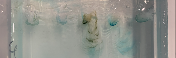
- After the experiment, both boxes' DNA strands were visible under regular light and UV light; however, instead of forming the traditional rectangles, they resembled magnetic field lines, like the shape of a bracket. Example: ) )) ) ))) )
Possible error
- When making the DNA samples for both boxes, we did make enough DNA samples for only one box instead of two, this could be why possibly the strands were hard to see.
Box 2
-
I confirmed that the electrical current was running properly when a fruit fly flew into the running buffer, flew back out into the air, and died once it had flown away from the box.
-
DNA strands were slightly more visible due to the dyeing of the DNA itself
Box 1
- DNA strands were less viable, but more strands were visible than in box 2; this is probably due to the dyeing of the running buffer instead of the DNA itself.
Attempt 4 & 5
For my fourth and fifth attempts, I made both boxes the same except for the amount of dye I used in each two drops of box one and one drop for box 2. I decided to dye the gel with methylene blue dye overnight while letting the gel set/harden instead of dying the DNA and Running buffer. I also switched the cherry sample out with a kiwi.
- All the fruit strained easier when doubling the fruit and buffer than in other previous attempts. Kiwi and blueberry produced the most juice out of the 5.
- Blueberries and banana DNA were found in a gel mixture instead of white strands like strawberries. Possibly the fruit's sugar in a gelatine form, forming around the DNA?
- Five minutes into running the experiment, one of the DNA bars/strands/molecules was visible in both boxes 1 and 2. It was rectangular instead of the shape of an electromagnetic field line.
- Boxes had 10x more visible DNA strands than other previous attempts. On the 4th & 5th attempts, it first appeared to be separating the molecules in a rectangular shape, but after the experiment, they were still simillar to an electromagnetic field line. Each DNA didn't have to distinctly of a unique pattern, but all were still different when compared closely.
- All fruit had partially visible DNA lines. Visible under regular light and UV light.
Amount of DNA extracted.
- Kiwi - DNA 1ml - water 1ml
- Bananna - DNA 3ml - water 3ml
- Strawberry - DNA 2ml - water 2ml
- mango - DNA1ml
- Blueberry- DNA 3ml - water 3ml
Possible error:
- Batteries died halfway through the experiment on both box and box 2
Attempt 6 & 7
I tested agar agar for attempts 6 and 7; however, since scientific grade agar agar is highly costly, I sought culinary grade agar agar. Other than the gel, I kept everything else in the experiment the same as my somewhat successful previous attempts. Including the same DNA extraction procedure and dying the gel instead of the DNA or running buffer.
- The culinary grade agar agar formed an estimated 7x harder than the pure gelatin. After researching some more, most chefs using culinary agar agar dilute it to avoid its hard and firm texture.
- The DNA samples were unable to move through the gel at all. I believe that this is possibly because of the thickness and firmness of the gel. I also believe that because the gel is so hard, its pores may have been more closed or too small that any visible DNA molecules, even with dye, could be seen.
Observations in all experiments
- Bubbles in strawberries and blueberries, throughout all the attempts, when extracting blueberries and strawberry DNA, they were the only fruits that seemed to have bubbles in the fruit juice during the isopropanol step.
- All frozen fruit samples did not show visible DNA during the extraction. Because of this, the frozen fruits did not generate any DNA to be used in the gel electrophoresis processes.
Analysis
Throughout my multiple experiments, I was able to collect five partially identifiable DNA samples throughout seven attempts. Throughout these attempts, I tried many different approaches to gel electrophoresis. During the trials, I tried different fruit, gel formulas, and staining methods to see which combination of the different methods worked the most effectively.
On my first attempt, I followed the regular procedure for DNA extraction, which was using a ziplock and detergent buffer and the normal creation of the gel electrophoresis box. However, on this first attempt, I stained the gel overnight with blue methylene dye. I also attempted using a mix of frozen and non-frozen fruits such as Bananas, strawberry, frozen corn, frozen peas and spinach.
On my second and third attempts, I followed the previous extraction and gel electrophoresis box procedure as completed in attempt one. On my third and second attempts, however, I switched the fruit that was unable to create the DNA strands with other possible fruits that could hopefully generate DNA strands. I ran two machines at the same time as well to make the trials more efficient. The only thing I changed between the two boxes and the first attempt was, for the 2nd box, I dyed the running buffer, and for the 1st box, I dyed the DNA after extraction, and it dissolved.
On my fourth and fifth attempts, I followed the previous extraction and gel electrophoresis box procedure as completed in attempt one. I made both boxes the same except for the amount of dye I used in each box gel instead of buffer or DNA. Two drops of blue methylene dye in box one and one drop in box 2. I also switched the cherry sample out with a kiwi.
On my sixth and seventh attempts, I followed the previous extraction and gel electrophoresis box procedure as completed in attempt one. I tested agar agar for attempts 6 and 7; however, since scientific grade agar agar is highly costly, I sought culinary grade agar agar, which ultimately did not work.
In conclusion attempt one, six and seven did not work, however I was able to collect slighty identifiable samples from attempt 2,3,4 and 5. I was able to collect samples of Banana and strawberry from attempts 2 and 3 and blueberry mango and kiwi from attempts 5 and 4.
Conclusion
In conclusion, for my experimental project, I was testing if it was possible to solve a fake fruit crime scene using gel elctrophoresis on a low budget, at home, and in other words, trying to see if it was possible to perform gel elctrophoresis with homemade gel electrophoresis elements. I strongly believed before my experiment that "If the homemade gel electrophoresis machine and elements managed to successfully separate the DNA molecules on the extracted fruit DNA, I believe that then the different suspect's DNA would be comparable to the criminal DNA. This will happen Because Gel electrophoresis uses the negative charge of DNA to separate the DNA's molecules based on their size, making the DNA differentiable to the human eye. This will ultimately lead to the conviction of the guilty fruit." - Anna McLellan. To prove my hypothesis correct and convict the guilty fruit. It took several trials. During two weeks, I was successfully able to collect 5 DNA samples. However, some bands faded and some DNA samples leaked from the wells. But, despite the errors, there was a successful match of the blueberry's DNA to the crime scene DNA (evidence), proving that the blueberry was indeed the murderer of the banana. Overall, I indeed did prove that my hypothesis was partially correct, and that it is possible to perform gel electrophoresis with homemade equipment (with a few errors.)
Application
This research on at-home gel elctrophoresis I hypothesis could be used in a school environment. Shockingly, many high schools now do not do gel electrophoresis labs to help their students due to the high cost of professional gel elctrophoresis elements. However, many high schools are now researching how to create a cost-effective and budget-friendly gel elctrophoresis lab for their students, but despite this, many high schools still do not do gel electrophoresis labs. This research could be used as a base platform for teachers and students alike to learn and improve on the low price method of this experiment.
Sources Of Error
Some sources of error during this experiment that could be improved upon for further research and more attempts were:
- All of the frozen fruit samples did not show visible DNA during the extraction. The frozen fruits were unable to isolate and have their DNA precipitated during the DNA extraction procedure. Because of this no samples were collected from the frozen fruits.
- The culinary grade agar formed an estimated 7x harder consistency than the pure gelatin. After researching some more, most chefs using culinary agar dilute it to avoid its hard and firm texture. Using this agar agar was unsuccessful. I hypothesize that because the agar agar was so thick the pores of the gel may have been too small and tight together, for any separation to occur.
- Not measuring correctly the amount of DNA and distilled water to dilute the DNA. During the processes of creating samples the weighing scale proved to be unreliable, giving wrong mesurments for the first experiment. Because of this error, attempts were not completed at the full capability of the trial.
- Batteries ran out of voltage halfway through attempts 7 and 6. Because of this, the attempt was not completed at its full potential. The batteries running out halfway through the experiment caused the experiment to end abruptly, Dueto this the DNA was not fully separated. Despite that, the trial was still somewhat successful.
These sources of error can be easily avoidable in future attempts.
Citations
Professor Drew Collop. (2020, September 25). Agarose Gel Electrophoresis [Video]. YouTube. https://www.youtube.com/watch?v=M1zFRf3fbQA
Synonyms and Antonyms of Words | Thesaurus.com. (2025, January 29). Thesaurus.com. https://www.thesaurus.com/browse/ideally
What is Forensic Science – Canadian Society of Forensic Science. (n.d.). Canadian Society of Forensic Science. https://www.csfs.ca/student-zone/student-zone/
EDVOTEK. (2021). Left at the scene of the crime: Introduction to forensic science. In EDVOTEK WORKSHOP. https://www.edvotek.com/site/pdf/Edvotek_Left_at_the_Scene_Booklet.pdf
Dictionary.com | Meanings & Definitions of English Words. (2025). In Dictionary.com. https://www.dictionary.com/
ELEMENTS OF AN EXPERIMENTAL PROJECT (pp. 1–1). (2009). https://www.cysf.org/wp-content/uploads/Experimental-Projects.pdf
Amoeba Sisters. (2017, September 27). Gel electrophoresis [Video]. YouTube. https://www.youtube.com/watch?v=ZDZUAleWX78
Gel electrophoresis. (n.d.). https://learn.genetics.utah.edu/content/labs/gel/
DNA extraction. (n.d.). https://learn.genetics.utah.edu/content/labs/extraction/
DNA Extraction Lab: Strawberry. (n.d.). https://www2.nau.edu/lrm22/lessons/dna_extraction/strawberry_dna.html#:~:text=We%20will%20use%20an%20extraction,the%20DNA%20inside%20the%20cells.
Khan Academy. (n.d.). https://en.khanacademy.org/science/up-class-12-physics/x0958a876c1afdc76:electric-charged-and-fields/x0958a876c1afdc76:electric-charge/a/electric-charge-ap1
Why is DNA negatively charged? (n.d.). https://byjus.com/biology/why-is-dna-negatively-charged/#:~:text=The%20phosphate%20backbone%20of%20DNA,DNA%20to%20be%20negatively%20charged.
Gel electrophoresis. (n.d.-b). https://www.sciencedirect.com/topics/agricultural-and-biological-sciences/gel-electrophoresis
Oxford Languages and Google - English | Oxford Languages. (2024, January 16). https://languages.oup.com/google-dictionary-en/
Citaition Generator. (n.d.). Scribbr. https://www.scribbr.com/citation/generator/folders/3YHBstiQCUIvklusVleaG/lists/1MT3PvlXiqprAUBo9rOeET/
Grammarly, Inc. (n.d.). *Grammarly* [Writing enhancement software for spelling and grammar]. https://www.grammarly.com
Gel electrophoresis - Labster. (n.d.). https://theory.labster.com/gel_electrophoresis_ag/
Lan, V. T. T., Loan, P. T. T., Duong, P. a. T., Thanh, L. T., Ha, N. T., & Thuan, T. B. (2012). Straightforward procedure for laboratory production of DNA ladder. Journal of Nucleic Acids, 2012, 1–4. https://doi.org/10.1155/2012/254630
Polymerase chain Reaction (PCR). (n.d.). Genome.gov. https://www.genome.gov/genetics-glossary/Polymerase-Chain-Reaction-PCR
Restriction Fragment Length Polymorphism (RFLP). (n.d.). Genome.gov. https://www.genome.gov/genetics-glossary/Restriction-Fragment-Length-Polymorphism-RFLP#:~:text=Restriction%20fragment%20length%20polymorphism%20(abbreviated,DNA%20with%20a%20restriction%20enzyme.
What is gel electrophoresis? (n.d.). https://www.yourgenome.org/theme/what-is-gel-electrophoresis/#:~:text=Gel%20electrophoresis%20is%20a%20technique,arranged%20in%20order%20of%20size.
Acknowledgement
Ms. Phillips, a teacher at The Calgary Girls Charter School. Thank you for overviewing my project and helping me stay on track.
Ms. Goday, a teacher at The Calgary Girls Charter School. Thank you for overviewing my project and helping me stay on track.
Emily N, Astrophysicist and Science High School teacher. Thank you, Emily N, for providing information on gel electrophoresis and your insight for the experiment.
Marian Alzubaidy, is a student at The Calgary Girls School. Thank you for reading over my paragraphs.