
From Pixels to Diagnosis: Neural Network-based Malaria Detection
Parth Sheth
Grade 11
Presentation
No video provided
Problem
Malaria: A Global Health Crisis
In the field of global health, malaria is a dangerous foe that has a devastating impact on both human life expectancy and socioeconomic advancement globally. Malaria is still a serious hazard, especially in areas like sub-Saharan Africa, South Asia, and portions of Central and South America, where there are an estimated 200 million cases and 400,000 fatalities each year. But beneath these sobering figures is a human tragedy of enormous proportions.
Malaria is a disease that hits people of all ages and socioeconomic status randomly in endemic areas. Pregnant women and children under five are especially at risk, as malaria contributes to high rates of morbidity and mortality in these populations. In addition to causing physical pain, the sickness has a significant financial cost since it reduces household resources, hinders productivity, and feeds the cycle of poverty and illness.
Furthermore, malaria has a significant social and economic impact in addition to its acute health effects. Families are frequently compelled to pay for treatment and care in endemic areas, where resources may be few and access to healthcare may be restricted, which exacerbates poverty and inequality. Furthermore, communities may become trapped in a cycle of poverty and poor health due to the long-term impacts of malaria on cognitive development and educational attainment.
Malaria not only has a negative impact on people's health and well-being but also presents serious difficulties for economies and healthcare systems. Budgets and resources for healthcare are already overextended, and the costs of treatment, prevention, and control initiatives are high. Furthermore, the indirect costs of malaria, such as missed productivity and slower economic growth, increase the disease's financial impact even more, especially in endemic areas where resources are already limited.
The Urgency of Early Diagnosis
Given the devastating effects of malaria on both people and the economy, early detection becomes very important in the fight against the illness. In addition to reducing personal suffering, prompt diagnosis and treatment of malaria cases help stop the disease from spreading throughout communities and lower the risk of serious complications and death.
However, there are several obstacles in the way of fast and accurate malaria diagnosis at the moment, making progress difficult. The detection and identification of Plasmodium parasites in blood smears is dependent on the proficiency of trained microscopists, and manual microscopy has long been regarded as the gold standard for diagnosing malaria. Although this method is praised for being inexpensive and flexible, it has some serious shortcomings, including as low throughput rates and a vulnerability to bias and human mistakes.
The use of manual microscopy presents substantial logistical issues in resource-constrained contexts where healthcare infrastructure may be inadequate. This hinders efforts to expand diagnostic services and reach underserved people. Furthermore, the possibility of unpredictability and inconsistency in diagnosis is introduced by the subjective character of human interpretation, further jeopardizing the accuracy and dependability of test results.
Moreover, manual microscopy takes time and expertise, which limits the pace at which diagnoses may be made. This delays the start of treatment and lets the condition worsen unabated. These delays can have disastrous effects, worsening the outbreak's severity and impeding efforts to limit transmission in areas with high malaria burdens where every second counts in the fight against the illness.
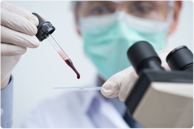
The image above is what the blood smear diagnosis method looks like.
A Novel Approach: Neural Network Diagnosis
Given these difficulties, I have imagined a unique method for diagnosing malaria that makes use of machine learning and artificial intelligence. Motivated by the latest developments in deep learning, I suggest creating a neural network-based diagnostic system that can quickly and precisely identify malaria parasites in blood smears.
Convolutional neural networks (CNNs), a family of deep learning algorithms created especially for image identification problems, are the foundation of this methodology. These neural networks may be taught to automatically detect and classify malaria parasites with previously unheard-of speed and accuracy by utilizing enormous datasets of annotated blood smear images.
Furthermore, there are a number of notable benefits to using neural network-based diagnostic systems as opposed to conventional microscopy techniques. First of all, by automating the diagnostic procedure, these systems can drastically cut down on the amount of time and experience needed to diagnose malaria, allowing for the quick screening of a large number of patients and the promotion of early illness detection and treatment.
Second, human error and prejudice inherent in manual microscopy are eliminated by the inherent objectivity and consistency of neural network-based diagnostic solutions. This improves the accuracy and consistency of diagnosing malaria by guaranteeing that test results are dependable and repeatable, independent of the test operator.
Neural network-based diagnostic systems are also highly scalable, which makes them ideal for implementation in environments with limited resources, such as those with restricted access to skilled workers and specialized equipment. The most vulnerable populations could benefit from rapid and accurate malaria detection by having neural network-based diagnostic software installed on portable, reasonably priced digital microscopy devices that are placed in rural and underprivileged locations.
To sum up, the creation of neural network-based diagnostic tools is a promising area of research in the fight against malaria, with the potential to completely change how the disease is detected and treated. In the battle against this terrible illness, we can increase test findings' accuracy and dependability, diagnose patients more quickly, and ultimately save lives by utilizing artificial intelligence and machine learning.
Method
Data Collection
Obtaining an appropriate dataset for the project's training and validation of the suggested neural network-based diagnostic system was an essential initial step. An excellent option was the Broad Bioimage Benchmark Collection (BBBC041), which provides an extensive collection of excellent microscope pictures that have been specially selected for use in biomedical image processing applications. There were three separate image sets in this dataset, for a total of 1364 photos. The dataset included images with a class label identifying the type of cell (red blood cell, gametocyte, ring, trophozoite, or schizont; the last four are infected cells) and a set of bounding box coordinates defining the spatial extent of each cell in the image. This is a noteworthy feature of the dataset as having prelabeled/annotated images greatly speeds up the overall process of creating a neural network.
Image Segmentation
After obtaining the dataset, the subsequent phase entailed training a convolutional neural network (CNN) to perform image segmentation, with the particular objective of identifying individual cells in microscopy pictures. Given that subsequent cell classification tasks depend on the precise identification of cell borders, image segmentation is a key preprocessing step in the diagnostic pipeline. Using the BBBC041 dataset's annotated bounding box coordinates, a supervised learning technique was used to train the CNN to identify the spatial characteristics that correspond to the locations of the cells in the photos. The CNN gradually learned to correctly identify cell boundaries by iteratively adjusting the network parameters using gradient descent-based optimization algorithms. This allowed the CNN to successfully segment the images into discrete sections that correspond to individual cells.
Cell Identification
After the picture segmentation model was trained and proven to work, the focus shifted to training a different CNN for cell identification. Cell identification entails assigning each segmented cell to one of the preset categories (i.e., red blood cell, gametocyte, ring, trophozoite, or schizont), in contrast to image segmentation, which focuses on defining cell borders. The CNN was trained using a supervised learning methodology to discover the discriminative characteristics connected to each cell type by utilizing the class labels supplied in the BBBC041 dataset. Through the process of utilizing backpropagation to optimize network parameters and presenting the segmented cell images as input, the CNN gradually acquired the ability to distinguish between various cell types according to their visual attributes. Iterative training and validation cycles allowed the CNN to identify cells with high accuracy and dependability.
Overall Diagnosis
The last stage in the diagnostic process entailed combining the picture segmentation and cell identification CNNs, each of which had been trained and verified separately, to carry out the comprehensive diagnosis of malaria infection. To do this, microscope pictures were processed using an image segmentation CNN to identify individual cells. Then, the segmented cell images were sent through a cell identification CNN to categorize each individual cell into a predetermined group. For every input image, the procedure produced a thorough diagnosis that showed the existence and quantity of various cell types linked to malaria infection.
This thorough methodological framework, which uses deep learning and convolutional neural networks to automate and expedite the diagnostic procedure, offers a fresh and promising way to diagnose malaria. In the fight against malaria, the suggested diagnostic method has the potential to improve patient outcomes, decrease turnaround times, and increase diagnostic accuracy.
Data:
I have finished the image segmentation process, but the cell identification neural network still needs to be completed training. The training took longer than expected and as such, I cannot report the accuracy and speed of the overall model yet. I will present it in-person at the CYSF.
Analysis
I have finished the image segmentation process, but the cell identification neural network still needs to be completed training. The training took longer than expected and as such, I cannot report the accuracy and speed of the overall model yet. This will be presented in-person at the CYSF.
Conclusion
I have finished the image segmentation process, but the cell identification neural network still needs to be completed training. The training took longer than expected and as such, I cannot report the accuracy and speed of the overall model yet. This will be presented in-person at the CYSF.
Citations
https://bbbc.broadinstitute.org/BBBC041
https://medium.com/@fractal.ai/guide-to-build-faster-rcnn-in-pytorch-42d47cb0ecd3
https://pytorch.org/tutorials/intermediate/torchvision_tutorial.html
https://www.cdc.gov/dpdx/malaria/index.html
https://www.cdc.gov/malaria/diagnosis_treatment/diagnosis.html#:~:text=Malaria%20parasites%20can%20be%20identified,the%20parasites%20a%20distinctive%20appearance.
The links above were the main sources used in my project, but small pieces of information from dozens of other websites were also used.
Acknowledgement
Thank you to my parents who supported me during the duration of this project and to my CYSF Coordinator Mr. Buhler for giving me the opportunity to participate.