The Interaction of Internal Health and External Triggers in Atopic Dermatitis
Siya Shetty
North Trail High School
Grade 11
Presentation
No video provided
Problem
What is Atopic Dermatitis?
Atopic Dermatitis, more commonly known as Eczema is a chronic skin condition that affects over 16.5 million individuals in the United States. Overall, about 31.6 million people in the United States have some type of Eczema. This particular skin condition is not curable but can be treated by various ways and prevention. There are many different types of Eczema including Atopic Dermatitis, Seborrheic Dermatitis, Contact Dermatitis, Nummular Eczema and Dyshidrotic Eczema. The most prevalent type of Eczema is Atopic Dermatitis and it often results in dry and inflamed skin. All variations of Eczema are characterized by redness, intense itching and inflammation. The National Eczema Association defines Eczema as a skin condition that causes itchiness, dry skin, rashes, patches and blisters. While Atopic Dermatitis isn't a life threatening skin condition, it is highly prevalent in children and can have both physical and emotional impacts. Rady's Children's Health specifies many of the effects of Atopic Dermatitis on small children.
Physical Impacts of Eczema
- Some physical effects of Eczema include inflamed, itchy rashes that could potentially become infected. Scratching the patches of Eczema often leads to complications because of fungal, bacterial or viral infections.
- Eczema is physically uncomfortable and cause disturbances at night. Many individuals report disturbances in sleep and daily activities such as sports, school and work relationships.
- Personally, Atopic Dermatitis caused irritation, burning and a constant severe itching sensation and this often disrupted my sleep.
Emotional Impacts of Eczema
- Emotional stress impacts both individuals with Eczema and their family members
- Eczema can cause loneliness, lack of confidence and social isolation because many individuals with Eczema are viewed as unhygienic and people often step back in fear. I personally relate to this emotional aspect because my Eczema impacted my social confidence and I often feared showing my arms or hands due to the red and inflamed wounds
- Depression and anxiety are common impacts of Eczema on individuals.
Significance
Many individuals with Atopic Dermatitis are aware of some symptoms and triggers but, many don't know the exact cause due to the fact that Eczema is often a combination of genetics, environmental triggers, skin barrier dysfunction and immune system sensitivity. I often wondered what triggered my eczema and why it often flared at the most unexpected times. What external and internal factors impacted my Atopic Dermatitis? How is normal healthy skin different from sensitive skin with Atopic Dermatitis? These are questions that many individuals fail to answer because Atopic Dermatitis doesn't have one exact cause. It has many causes that are difficult to grasp and understand.
Research Focus
This research project aims to explain some of the possible causes of Eczema and their interactions. Immune sensitivity, skin barrier dysfunction and overall gut health are internal health factors that interact with environmental and lifestyle related triggers. These impact the severity of an individual's Atopic Dermatitis. I will also be collecting data from students in my school related to their personal experiences with Eczema. I predict that students will have varying experiences with eczema and trigger. This is because eczema is a complex skin issue which involves several factors and the genetics of an individual.
Method
My non experimental research project consists of four major segments. These are :
- Background Research
- Detailed research about external triggers and internal health factors
- Data from students, dermatologists and personal experiences
- Analysis into the interaction between these external and internal factors in Atopic Dermatitis
Reliable sources were used that provided research on the particular external and internal factors that were to be analyzed. Each source focused on particular areas such as immune sensitivity, skin barrier dysfunction and gut health.
To account for the variation in symptoms, a few students were surveyed at my school and they shared their personal experiences with Eczema including their observations on triggers. I also utilized a table of observations from my personal experience with eczema in India and how the external factors there influenced the severity.
Background Research
This particular segment of my research project digs deeper into the general system of the skin and how the skin works. This section provides general information about the immune system's role in the skin and the structure of the layers.
External Triggers and Internal Health Factors
This portion of the project dives deeper into each of the factors including immune sensitivity, skin barrier dysfunction and gut health as well as environmental and lifestyle related external triggers. This helped me gain a more detailed understanding of each of the factors and how they impact the skin. This information also helped me analyze the link the interactions between these factors. I used AI to clarify some complex terminology. Then, I reread the scientific research papers and developed a comprehensive understanding. From there I created understandable and detailed explanations using my own words while still keeping important medical terminology.
Analysis : I utilized the research I had gathered to form connections between external and internal factors that impact Atopic Dermatitis. I highlighted the interactions between these factors and how Eczema is often triggered by the combination of the two.
Data
I collected data from students at my school to account for the variations in symptoms. Atopic Dermatitis affects each individual uniquely and each person has specific triggers. Collecting anecdotal data helped me learn about what external triggers affected individuals of my age. I also made a table of observations from my personal experiences with eczema in my summer 2025 India Trip. This trip made me realize the different external factors that increased the severity of my Eczema.
Interpretation
I will be using the research I gathered to create an understandable and interactive model for my physical presentation about the different parts of the immune system and the factors which impact the severity of Atopic Dermatitis. My model will include immune cells, skin layers and triggers to show a comprehensive understanding of eczema.
Research
Background Research
The skin is the heaviest and largest organ in the human body and acts as a central sensory organ. The skin performs several crucial tasks like protecting the body from pathogens, toxic substances, the cold and sun rays. In addition to protection, the skin also plays a role in regulating body temperature, storing water, fat and metabolic products. The skin is composed of 3 main layers each with a specific function. These layers are called the Epidermis, Dermis and Subcutis.
Epidermis
The epidermis is the outermost layer of the skin and varies in thickness depending on the part of the body. The role of the epidermis is to provide an effective barrier that separates the individual from the surrounding environment. It is primarily composed of keratinocytes which are cells that produce keratin. These cells establish a protective barrier, preserve moisture and prevent pathogens from entering the skin. This layer of the skin is constantly renewing itself through shedding. New cells are built in the lower layers of the epidermis. As these cells reach the surface, they die and shed, resulting in a continuous renewal cycle.
The epidermis also consists of other significant cells like merkel cells, lymphocytes, langerhans and melanocytes. Lymphocytes and langerhans play an important role in protecting the body against pathogens while melanocytes are involved in pigmentation as they produce melanin. Merkel cells are nerve cells that allow one to sense pressure.
Dermis
The Dermis is the middle layer of the skin that is made up of a network of collagen fibres. These fibres allow the skin to be strong as well as elastic. The Dermis also consists of a network of capillaries. Capillaries are the body's smallest blood vessels that deliver nutrients and oxygen to surrounding cells. This layer is also involved in temperature regulation, helping your body to cool down or preserve heat. This happens because of the blood vessels in the Dermis that use the processes of vasodilation and vasoconstriction to regulate body temperature.
Other Key Components of the Dermis Include
- Sebaceous glands : These glands produce sebum which is an oily substance that lubricates the skin and keeps it hydrated.
- Sweat Glands : Sweat glands produce sweat when an individual is too hot or stressed. Sweat glands also help to regulate body temperature.
- Hair Follicles : This layer of the skin contains hair follicles which produce hair for your body.
- Sensory Cells : Detect pressure and touch.
Subcutis (Hypodermis)
The Subcutis is mostly made up of fat and connective tissue. Just like the Dermis, the Subcutis contains blood vessels and nerves. Between the Dermis and the Subcutis are tiny cavities that contain a tissue composed of fat and water. This fat layer protects the bones, organs and other key structural components from injuries and sudden shock. Overall, the Subcutis has the primary role of protecting the skeletal system, organs and muscles from harm.
Other Key Components of the Subcutis Include
- Macrophages : These are a type of white blood cell that use the process of phagocytosis to engulf foreign pathogens and present the antigens to alert other cells involved in immunity and protection.
- Lymphatic Vessels : These are a network of vessels that are closely associated with the circulatory system. They help to regulate fluid balance and contain a fluid called lymph. The lymphatic system also contains different types of white blood cells like T Lymphocytes, B Lymphocytes, Macrophages and Monocytes.
- Nerves : Nerves on this layer send out electric signals and are responsible for the transfer of information to and from cells, organs and muscles.
- Fibroblasts : This is a type of connective tissue cell that produces collagen.

This image depicts the three layers of the skin and illustrates a general overview of the skin. Retrieved from : Fld. (2025, February 26). Understanding the layers of your skin. Florida Dermatology and Skin Cancer Center. https://fldscc.com/understanding-the-layers-of-your-skin/
Background Research - Analysis of the Immune System
The skin is a layer of protective barrier that guards your internal body from foreign pathogens. To achieve this. the skin harbours several types of immune cells that are involved in innate and adaptive immune response. Innate immune response refers to parts of the organism that are involved in non specific defence that creates a protective barrier. Adaptive immune response is specialized and more targeted towards specific pathogens. The skin itself is part of the innate immune system because it is one of the first lines of defence. The skin has a complex immune system that is represented by Skin Associated Lymphoid Tissue (SALT). The SALT system consists of several cells like dendritic cells, mast cells, B lymphocytes, T lymphocytes and keratinocytes. This system also protects against foreign pathogens and plays a role in inflammatory diseases like autoimmune and hypersensitivity disorders. The Epidermis contains three major types of immune cells including keratinocytes, melanocytes and langerhans cells. The Epidermis itself has four layers ranging from the deepest to the top layers : the Stratum Basale, Stratum Spinosum, Stratum Granulosum and Stratum Corneum.
Stratum Basale
- This layer consists of non-specialized cells which are constantly involved in dividing into new cells. This process is known as mitotic activity.
Stratum Spinosum
- In this layer, the non-specialized cells from the Stratum Basale become specialized into cells which produce keratin. Keratin helps to support your Epidermis, heal wounds and provide protection.
Stratum Granulosum
- It is composed of keratinocytes which produce keratin forming proteins as well as lipids. These are crucial for forming the skin's protective barrier because keratin provides protection. Lipids play a key role as a protective barrier and they also prevent sun damage and moisture loss.
Stratum Corneum
- This layer contains fully matured keratinocytes. They help protect the individual by blocking potentially toxic pathogens. These cells also maintain stable water and electrolyte levels which is known as hydroelectrolytic homeostasis.
As addressed earlier, the Epidermis also consists of other cells such as T lymphocytes, melanocytes and Merkel cells. The Stratum Basale contains melanocytes which protect DNA from sun damage by producing a pigment called melanin. Merkel cells are responsible for feelings of touch throughout the Epidermis and, the Basale and Corneum layers contain T lymphocytes which are involved in specific adaptive immunity. The middle layer of the skin, the Dermis consists of immune cells like dendritic Cells, T Lymphocytes, natural killer cells, mast cells, macrophages and neutrophils.
Background Research - Keratinocytes
These cells play an important role in maintaining the functions of the Epidermis and they also contribute to inflammatory processes by producing cytokines. Keratinocytes can recognize the difference between regular micro-organisms and foreign pathogens. They also recognize structures called Pathogen-Associated Molecular Patterns (PAMPs). These structures are important because they recognize several foreign pathogens. These PAMPs are usually composed of compounds like flagellins, nucleic acids. teichoic acids, lipopolysaccharides and much more. There are also molecules that attach themselves to PAMPs. These molecules are known as Pattern Recognition Receptors (PPRs) and include Toll-Like Receptors (TTRs). The binding of PAMPs and the receptors are extremely important in adaptive and innate immunity. There are also other receptors like NLRs which are involved in the recognition of foreign substances like irritants and toxins. Keratinocytes also produce Antimicrobial Peptides (AMPs). These can perform several functions like eliminating pathogens and signalling other immune cells. Keratinocytes also activate T cells, B cells and dendritic cells. As shown, Keratinocytes play an important role in the immune system because they can recognize foreign pathogens through the use of NLRs, TTRs, and PAMPs. In conclusion, Keratinocytes are not just cells which produce protective Keratin. They are cells which play an active role in the body's immune system. I will be further researching about how the immune system changes in Eczema prone and sensitive skin.
Background Research - T Lymphocytes
The skin contains a T Lymphocyte population mainly composed of memory T cells. T lymphocytes can be found in the Stratum Basale of the Epidermis and in clusters around the capillaries in the Dermis. T cells participate in immune response and include several types such as Th1, Th2, Th3, Th17 and regulatory T cells (Tregs). These types of T cells have been found most particularly in individuals with infectious and inflammatory diseases. Th1 lymphocytes are involved in infections and autoimmune or immune mediated diseases like Psoriasis. Th2 lymphocytes participate in response to foreign pathogens in allergic diseases such as Atopic Dermatitis and Psoriasis. Th2 T cells also release various cytokines like IL-4, IL-5 and IL 13 in immune responses. Cytokines can be defined as proteins that help with the activity of immune cells. Th17 cells are also involved in the development of Psoriasis and Atopic Dermatitis. The regulatory T cells suppress immune response and ensure that healthy tissue isn't affected. In conclusion, T cells are immune cells which are involved in the immune response of the skin.
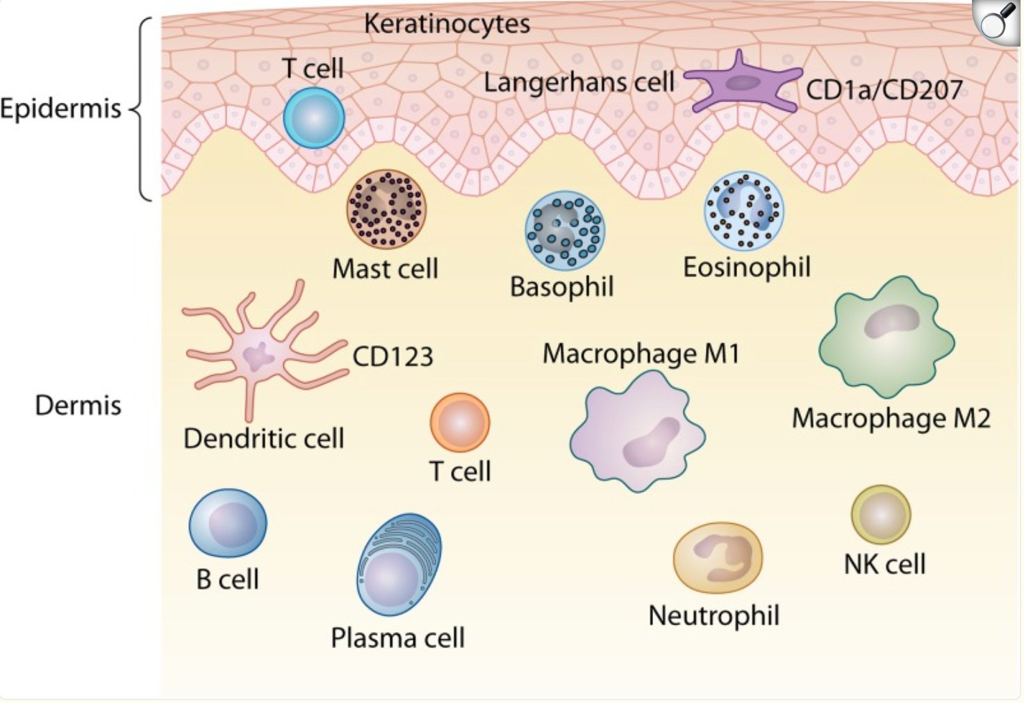 This image depicts the various different immune cells present in the layers of the skin and provide a general visualization. Retrieved from :
Quaresma, J. a. S. (2019c). Organization of the skin immune system and compartmentalized immune responses in infectious diseases. Clinical Microbiology Reviews, 32(4). https://doi.org/10.1128/cmr.00034-18
This image depicts the various different immune cells present in the layers of the skin and provide a general visualization. Retrieved from :
Quaresma, J. a. S. (2019c). Organization of the skin immune system and compartmentalized immune responses in infectious diseases. Clinical Microbiology Reviews, 32(4). https://doi.org/10.1128/cmr.00034-18
Internal Health Factors of Atopic Dermatitis
Skin Barrier Dysfunction
A normal skin barrier contains several proteins which help to maintain a normal barrier through regular function. Individuals with Atopic Dermatitis (AD) skin isn't as effective at keeping irritants and pathogens from entering the skin. Skin suffering from Atopic Dermatitis is also weak at controlling moisture. Eczemahelp.ca refers to the skin as a "brick-and-mortar wall" that prevents pathogens from entering, Eczema prone skin is more damaged which allows irritants to enter easily causing inflammation and moisture loss. In the Epidermis, there is a constant renewal of cells because the keratinocytes are constantly evolving. Keratinocytes migrate from the deepest layers of the Epidermis to the Stratum Corneum. Here, keratinocytes become corneocytes through the process of cornification. Corneocytes are keratinocytes which are dead and provide a strong barrier. During this cycle, keratinocytes produce barrier proteins like filaggrins (FLGs), loricrin, transglutaminases (TGs) and much more. Additionally, the AMPs that keratinocytes produce help maintain the skin homeostasis and kill harmful micro-organisms. The Stratum Corneum also contains three types of lipids which prevent waterloss. These include cholesterol, free fatty acids and ceramides.
The exact cause of Atopic Dermatitis is still not concluded because this involves several variables. For instance, the overexpression of Th2 and Th22 cytokines contribute to skin barrier dysfunction because it alters lipid and protein content. The overexpression of these cytokines impact essential proteins. For instance, FLG is a protein which helps maintain normal skin pH levels and lock in needed moisture. Atopic Dermatitis skin often lacks FLG and this changes the shape of corneocytes. This results in increased inflammation. The lack of FLG is caused by genetic factors, overexpression of cytokines and much more. The deficiency of FLG causes an individual to be less tolerable to irritants. FLG is often decreased by the overexpression of IL-4, IL-13, IL-25, IL-17A and IL-22 cytokines mostly from T lymphocytes.
Th2 cytokines also impacts the abundance of Corneodesmosin (CDSN). This is a intercellular protein which maintains a regular skin barrier. According to Significance of Skin Barrier Dysfunction by Kim and Leung (2018), a study shows that the lack of CDSN resulted in increased pathogenic entry in a skin model. The cytokines which contributed to this defect included several types like IL-4, IL-13, IL-22, IL-25 and IL-31. The AMPs produced by keratinocytes are also impacted by Th2 cytokines. They suppress the production of AMPs which leads to a weakened barrier that is more prone to infection. For instance, deficiency in AMPs led to more S.aureus infection. Staphylococcus aureus is a type of bacterium which is able to grow on AD skin because of a weakened skin barrier.
Additionally, skin with Atopic Dermatitis has a defected lipid barrier in the Stratum Corneum due to abnormalities in the expression of enzymes. These enzymes are usually involved in the synthesis of free fatty acids and ceramides for the lipid barrier. However, they are expressed differently in AD skin. This changes the lipid composition. It causes an abnormal skin barrier and increased permeability of the Stratum Corneum. In conclusion, Eczema prone skin is more susceptible to inflammation because Th2 over expressive cytokines weaken the barrier, and barrier defects further increase cytokine activity.
Internal Health Factor - Immune Sensitivity
The immune system plays a role in protecting the body from irritants by reacting to them when in contact. Individuals with Atopic Dermatitis have an overreactive immune system and this hypersensitivity results in eczema flares. Additionally, eczema is further amplified by the scratch-itch cycle. The need to scratch an eczema flare is triggered by signals from the nervous system. This worsens the region and makes it more prone to pathogenic entry. Increased pathogenic entry further amplifies immune activation in an already inflamed skin. Atopic Dermatitis is heavily impacted by immune mediated abnormalities in the skin. Often, genetic factors play a role in this. Immune sensitivity is also illustrated by increased levels of the antibody immunoglobulin E (IgE), Eosinophils and Tregs.
Regulatory T cells (Tregs)
Regulatory T cells (Tregs) impact immune balance through the process of suppressing immune responses. They regulate immune responses (like cytotoxic T cells) to ensure that healthy tissue isn't damaged. Many Tregs express CD25 and the transcription factor called Foxp3. These can be described as essential markers needed for Tregs to function normally. CD25 are also expressed by other T cells but it is used to recognize Tregs.
Individuals with Atopic Dermatitis (AD) skin are seen to have an abnormally function T cell population which transforms into an overreactive Th2 response. In some individuals, the Foxp3 gene goes through a rare mutation which results in a congenital disorder known as IPEX. This syndrome causes a deficiency in CD25+ Tregs. This results in immune imbalance. Individuals with this mutation may develop inflammatory conditions like airway inflammation and Eczema.
The Role of Regulatory T cells in Atopic Dermatitis by Agrawal, Wisniewski, and Woodfolk (2015) suggests that adults with Atopic Dermatitis actually have increased levels of Tregs which express CD25 and Foxp3. Now, this might seem odd but studies show that Tregs are not necessarily reduced in AD skin but they function abnormally. Individuals with AD have colonies of S.aureus and increased IgE antibodies. This might indirectly impact the ability of CD25+ Tregs to function normally. Tregs can also transform into different types of T cells. This is known as "plasticity". Tregs can turn into Th2 cells and Th17 cells. These are the same cells involved in the overreactions of the immune system. In conclusion, the abnormality of Tregs can impact the development of inflammatory conditions like Atopic Dermatitis. There is still further research to be done on Tregs and their role in Atopic Dermatitis because they possess complex properties.
Eosinophils
Eosinophils are a type of white blood cell (leukocyte) which helps defend the body against foreign pathogens and irritants. While they play a protective role, eosinophils have been observed to contribute to the development of inflammatory conditions. Individuals with Atopic Dermatitis generally have higher levels of eosinophils circulating in their blood. This higher concentration of eosinophils is referred to as Eosinophilia.
Eosinophils ultimately influence the number of cytokines released. For instance, in Asthma eosinophils are activated by cytokines from Th2 cells. Additionally, eosinophils release a high number of cytokines which results in inflammation. These cytokines lead to increased irritation and sensitivity in the skin. Overall, a higher level of eosinophils in individuals with Atopic Dermatitis impacts the hypersensitivity of the immune system.
Immunoglobulin E Antibody (IgE)
Immunoglobulin E (IgE) is suggested to play an important role in the development of inflammatory conditions like Atopic Dermatitis. IgE is an antibody which helps protect the body against foreign pathogens and infectious diseases through specific immune responses. IgE helps trap potentially harmful substances and alert the immune system. Individuals with Atopic Dermatitis (AD) skin often have high serum levels of IgE in their bodies. The term "serum level" refers to the amount of antibodies produced by the immune system. IgE contributes to the production of several cytokines such as IL-1, IL-3, IL-4, IL-5 and IL-6. Some of these cytokines such as IL-4 and IL-5 are released by overreactive Th2 cells which increases the sensitivity of the immune system. Increased serum levels if IgE contribute to a sensitive inflammatory response. Although, IgE antibodies are an important factor in Atopic Dermatitis, research is ongoing to fully understand its role in immune sensitivity.
Internal Health Factor - Atopic Dermatitis and Gut health
Atopic Dermatitis is a complex mix of several factors such as skin barrier dysfunction and immune sensitivity. However, the environment of the gut heavily influences the health of one's skin. Additionally, it also leads to immune sensitivity and skin barrier dysfunction. Patients with Atopic Dermatitis have been observed to lack a diverse microbial population in their gut compared to healthy individuals. The term microbiome refers to the variety of microorganisms situated on certain body parts. In the gut, a diverse population of beneficial microorganisms influence the development of important immune response cells such as Treg cells and the Foxp3 factor. They also play a role in the specializations of Th1, Th2 and Th17 cells. As known, Treg cells play an important role in regulating immune response and preventing overreaction. They regulate eosinophils, mast cells, basophils, IgE production and T cell specializations. Gut bacteria like Bifidobacterium, Lactobacillus and Clostridium release metabolic products such as butyric and propanoic acid. These products help activate and generate Treg Cells. Short Chain Fatty Acids (SCFA) possess anti-inflammatory effects and help keep the gut barrier intact.
The growth of microbes start as soon as a child is born. These microorganisms start developing and eventually mature by the time a child is 2 to 3 years old. One factor that influences the gut population includes the type of delivery. Vaginal delivery allows the child to become exposed to microbes from the mother. For instance, vaginal delivery exposes the baby to Bifdobacterium and Bacteroides. They help develop steady immune responses. In comparison, Cesarean section (C-section) babies become exposed to Streptococcus, Staphylococcus and C Difficile. The way a baby is fed during early childhood also impacts gut health and growth. Formula and breast feeding increases populations of Bifidobacterium. Bottle fed babies have higher concentrations of Ecoli and C Difficile. To conclude, experiences such as vaginal delivery, breast feeding, interactions with siblings and pets increase Tregs, microorganisms and SCFAs.
Individuals with Atopic Dermatitis (AD) skin have low biodiversity of microorganisms. As a result, this affects immune and inflammatory responses. It is important to note that patients with severe AD skin have less Bifidobacterium than individuals with mild AD. Additionally, AD patients have increased populations of Staphylococcus populations compared to healthy individuals. Microbiome of the Skin and Gut in Atopic Dermatitis by Eun Kim and Sung Kim suggests that the imbalance of gut bacteria and reduced SCFA leads to a "leaky gut." This results in the absorption of toxins, poorly digested food and microbes into systemic circulation. As it reaches the skin, Th2 response occurs resulting in tissue damage. Therefore, disruption of gut bacteria during early childhood can influence the development of Atopic Dermatitis.
External Environmental Factors of Atopic Dermatitis
Climate and Humidity
Many individuals with Atopic Dermatitis experience flares due to extreme hot and cold temperatures. For instance, the harsh winters of Northern North America can trigger and increase dryness and itchiness. In general, low humidity and temperatures increase skin barrier dysfunction and stress. Exposure to extreme cold temperatures contributes to the activation of cytokines which further increases sensitivity and the scratch-itch cycle. Cold conditions increase moisture loss through the epidermis from the inner body. This is known as Transepidermal Water Loss (TEWL) and this causes dryness. Extremely high temperatures also increase Atopic Dermatitis flares due to irritation, overheating, perspiration and bacterial growth. Low humidity dries out skin and makes it more susceptible to irritation. However, hot and humid conditions increase sweating and flare 0-4 days after exposure. Additionally, current environmental issues such as climate change contribute to worsening Atopic Dermatitis symptoms. Climate change driven events such as wildfires can cause sudden changes in temperature. These changes can increase flare-ups in patients who already have a compromised skin barrier.
Allergens and Pollutants
In urban areas, the risk of developing inflammatory like Atopic Dermatitis is increased due to emissions and air pollutants. Some molecular air pollutants which trigger Atopic Dermatitis include nitrogen dioxide, sulphur dioxide and benzene. Particulate matter from the combustion of fossil fuels also heavily impact the skin's reactivity. Such pollutants penetrate and irritate Atopic Dermatitis (AD) skin due to skin barrier dysfunction. For example, the dysfunction of the FLG proteins increases susceptibility to pollution. The pollutants irritate the skin and this results in an ongoing cycle. Components of indoor environments like renovations, paint and furniture can also cause flare-ups. The combination of outdoor pollutants, indoor components and allergens like dust mites increase severity of Eczema.
Additionally, climate change and air pollution are currently altering the pattern and composition of allergens like pollen. Individuals with inflammatory conditions are often sensitive to pollen as they activate IgE antibodies. Climate change impacts pollen season and concentration. For instance, warmer urban areas experience higher and earlier levels of pollen release.Air pollution like increased carbon dioxide levels can alter pollen proteins and increase allergenicity. Finally, climate change can promote growth of non-native plants with unfamiliar pollen.
Water Quality
Eczema can also be influenced by water quality, especially hard water. Domestic hard water refers to when minerals are dissolved in water from sedimentary rock filtration. Hard water often contains calcium through the presence of calcite and dolomite. The "hardness" of water can be measured by the concentrations of these minerals. In individuals with Atopic Dermatitis, hard water can penetrate the skin and cause dryness and increased barrier dysfunction. For instance, bathing in hard water with AD skin can increase the deposition of soaps such as sodium lauryl sulfate which cause Transepidermal Water Loss (TEWL). In conclusion, water quality could potentially increase the sensitivity of Atopic Dermatitis skin.
External Lifestyle Related Factors
Sleep
The irritation and severe itching associated with Atopic Dermatitis can have a profound impact on the ability to sleep properly. Atopic Dermatitis often worsens at night, disrupting sleep patterns which leads to worsening sleep quality. The worsening of Eczema at night is due to body temperature changes and moisture loss which increases an individual's tendency to itch. The itching then initiates the scratch-itch cycle. Eczema has also been observed to disrupt Rapid Eye Movement (REM) sleep. Reduced sleep quality poses several risks such as cognitive decline in areas of memory, attention and function.
Diet
In Atopic Dermatitis, the diet an individual follows can trigger flare-ups, IgE mediated sensitivity or late eczematous reactions. Allergic reactions could occur hours to days after the digestion of a certain food. Food allergies have mostly impacted infants and children with Atopic Dermatitis as they are most sensitive. Allergic reactions to ingredients such as wheat, nuts and soy can be described as health effects stemming from overreactive immune responses. These responses include IgE antibodies and non-IgE mediated responses. Immediate reactions are IgE mediated and can occur within minutes after digestion. These types of reactions worsen Atopic Dermatitis due to increased sensations of itchiness. Many patients have observed that the elimination of a diet related trigger helped improve Atopic Dermatitis. In comparison, late eczematous reactions could occur after hours or days. There is still further research to be done on late eczematous reactions and their impact on Atopic Dermatitis. In conclusion, an individual with Atopic Dermatitis could be sensitive to certain foods due to specific immune responses. These foods trigger reactions in several different ways.
Analysis
The research collected from scientific papers shows the potential correlations between external triggers and internal health factors of Atopic Dermatitis. Atopic Dermatitis is a complex skin condition which involves several factors and irritants. It is important to note that eczema does not occur due to one condition or factor. For instance, the family history of an individual could impact the stability of their immune system and skin barrier. Skin barrier dysfunction affects essential proteins such as AMPs and FLGs which help maintain skin pH and the integrity of the barrier. Abnormalities in the skin barrier also impact the rate of S.aureus infections and moisture loss. Additionally, the overreaction of Th cell cytokines further impair essential proteins. An overreactive immune system involves many components such as abnormalities in Treg cells, Eosinophilia and IgE antibodies. The gut microbiome of an individual influences the ability of the immune system to function properly. Gut microbiome has an impact on an individual since early childhood due to factors like type of delivery, sibling interactions, genetics and feeding methods. This shows how external triggers like hard water, climate and humidity can impact an already sensitive and inflammation prone skin. Allergens such as pollen, particulate matter and sulphur dioxide can penetrate quick and deep. Overall, this research indicates how external triggers and internal factors can dynamically impact the severity of Atopic Dermatitis flare-ups.
Data
Case Study : Anecdotal Data from Personal Experience
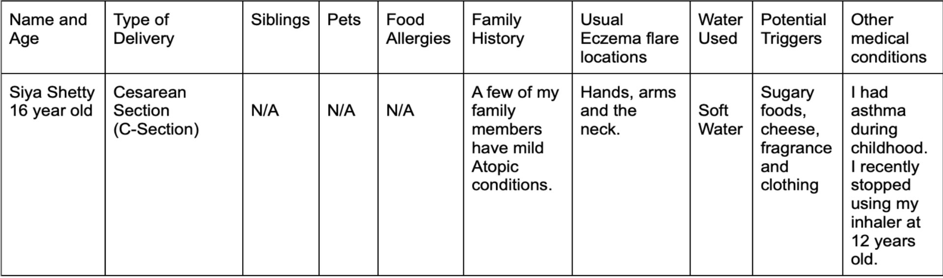
Recently during the summer of 2025, I visited India from early July to late August. My trip was about 2 months long in which I experienced changes in my eczema. My eczema became severe and gradually worsened during this period. The first data table provides important information about my health and how it relates to the pathogenesis of Atopic Dermatitis. The second data table provides my qualitative observations on my eczematous conditions.
General Atopic Dermatitis Data
Analysis
In the table above, I highlight the key factors and characteristics which may impact the severity of my Atopic Dermatitis. As shown by the data table, my type of delivery was C-section. This may have affected the development of beneficial gut bacteria during early childhood. Additionally, I have no interactions with siblings or animals. This indicates that I may have a decreased population of normally functioning Treg cells. Zero interactions with siblings or pets may have also impacted the amount of Short Chain Fatty Acids (SCFA) present in my body. This could have impacted my gut barrier and the systemic circulation of poorly digested food. Also, a few of my family members have milder Atopic conditions which could have genetically influenced the severity of my Atopic Dermatitis. I also use filtered soft water to ensure less irritation. It is important to note that after my asthma slowly recovered, my Eczema got more irritated. This may indicate a correlation between asthma and eczema.
Case Study : Personal Experience in India
Important Information
Water Used : Hard Water
Humidity Levels : High
Temperature : 26-32 degrees Celsius
Haircare : I brought Vichy's Dercos anti-dandruff shampoo to India instead of my other medicated Nizoral shampoo.
Personal Experience with Atopic Dermatitis in India
| Week | Observations |
|---|---|
| 1 |
|
| 2 |
|
| 3 |
|
| 4 |
|
| 5 |
|
| 6 |
|
| 7 |
|
| 8 |
|
Analysis
During my trip in India, my Atopic Dermatitis became increasingly inflamed and irritated over eight weeks. One of the main factors which impacted my eczema was the high levels of humidity in Mangalore. Mangalore is a city in the South Indian state of Karnataka and it embodies a tropical climate. The high temperatures mixed with high levels of humidity may have increased flare-ups. This is most likely due to increased sweating resulting in irritation. In Calgary, my Eczema becomes irritated during cold winters due to low humidity. However, the severity of my eczema was extremely low compared to my trip in Mangalore. This shows that my skin may be more sensitive to hotter and more humid climates. The water I used when showering in India was hard water. I predict that this factor made my eczema a lot worse because of the burning sensations. As the trip went by, the severity of my eczema gradually increased.
Emotional/Physical effects include :
- Burning Sensations
- Trouble sleeping
- Feeling "sick" after waking up
- Discomfort
Difference between Calgary and Mangalore's Environment
Mangalore
According to timeanddate.com, the average weather conditions ffrom 2012-2021 in Mangalore during the months of July and August include high temperatures and humidity.
July
The highest average temperature for July is 29 degrees celsius while the lowest temperature is around 23 degrees celsius. Precipitation : Average precipitation is about 884.6mm Humidity : The month of July is one of the most wettest and humid months of the year. The humidity at this time is usually 88%
August
The highest average temperature for August is once again 29 degrees celsius while the lowest temperature is also 23 degrees celsius. Precipitation : level of precipitation decreased during August to about 766.7mm Humidity : 88%
Calgary
According to timeanddate.com, the average weather conditions from 1992-2021 in Calgary during the months of July and August include high temperatures and low humidity
July
The highest average temperature during July is 24 degrees celsius while the lowest average temperature is 10 degrees celsius Precipitation : Level of precipitation is about 61.5mm Humidity : Humidity at this time is about 63%
August
The highest average temperature during August is 23 degrees celsius and the lowest average temperature is 9 degrees celsius. Precipitation : Precipitation drops to 50.5 mm Humidity : 61%
Analysis
This shows how generally higher levels of humidity and temperature may irritate the sensitive skin barriers of individuals with Atopic Dermatitis. Extreme temperatures may act as environmental triggers in Atopic Dermatitis.
Pollution and Air Quality In Mangalore and Calgary
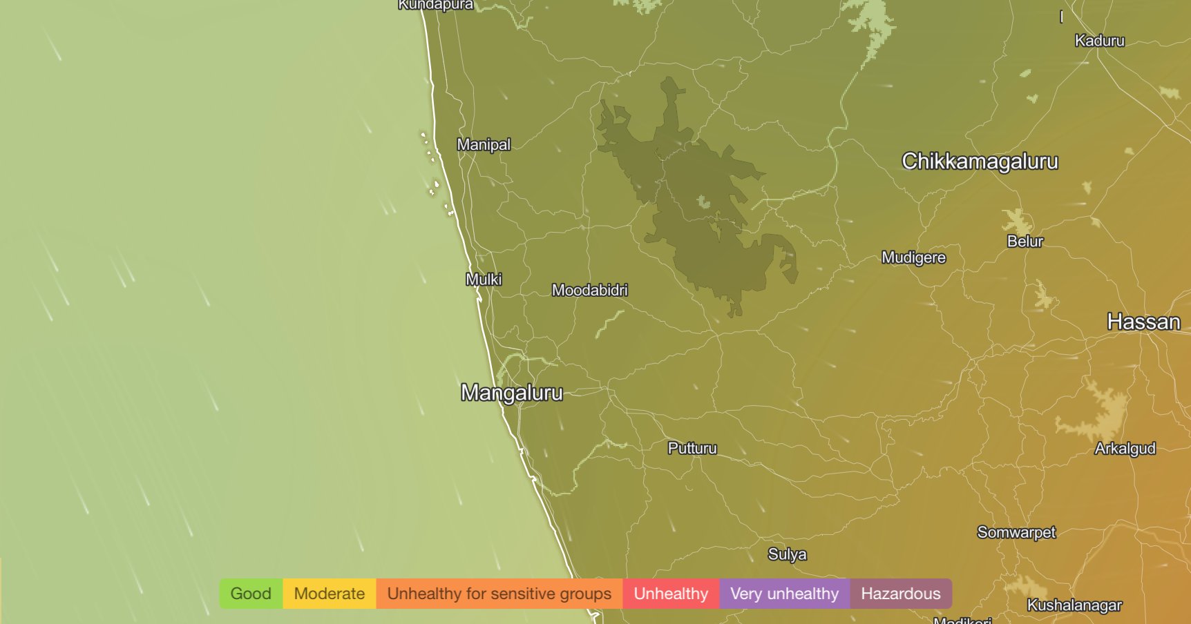
According the IQAir, the air pollution currently present in Mangalore is moderate at a rating of 77. The major pollutant in Mangalore is a type of particulate matter called PM2.5. The PM2.5 concentration in Mangalore is about 4.6 times greater than World Health Organization's (WHO) guideline.
Some Major Pollutants include
- Particulate Matter : PM2.5 at about 20 µg/m³
- Particulate Matter : PM10 at about 22 µg/m³
- carbon monoxide : Approximately 290 ppb (parts per billion)
- sulfur dioxide : approximately 2 ppb
- nitrogen dioxide : approximately 8 ppm
- ozone : approximately 22 ppb
The following air pollution map from IQAir illustrates the real time air quality conditions of Mangalore
 Retrieved from :
IQAir. (n.d.-b). Mangalore Air Quality Index (AQI) and India Air Pollution | IQAIR. https://www.iqair.com/ca/india/karnataka/mangalore
Retrieved from :
IQAir. (n.d.-b). Mangalore Air Quality Index (AQI) and India Air Pollution | IQAIR. https://www.iqair.com/ca/india/karnataka/mangalore
Calgary
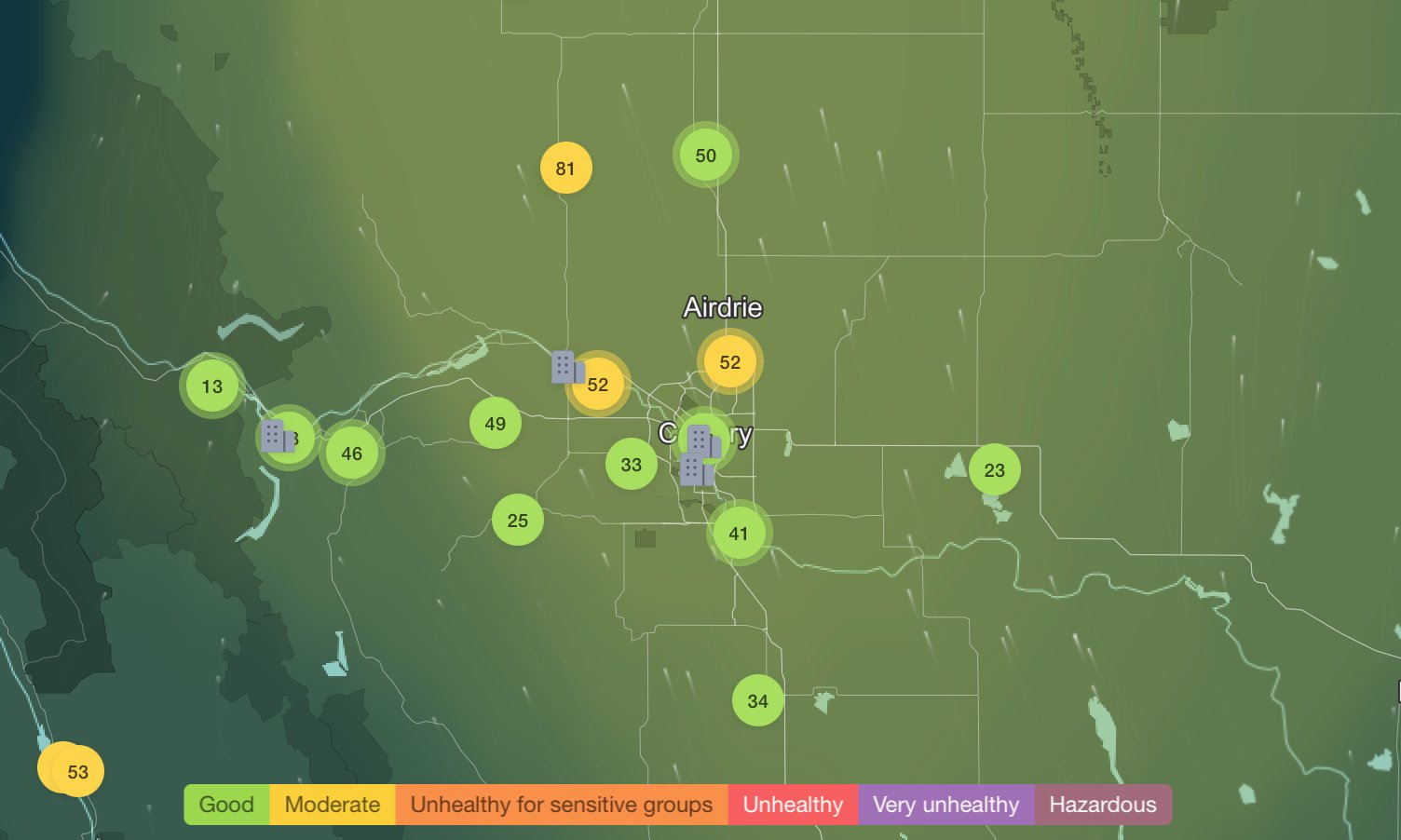
According to IQair, the air pollution currently present in Calgary is good at a rating of 44. The main pollutant in Calgary is also PM2.5. The PM2.5 concentration in Calgary is about 1.6 times greater than World Health Organization's (WHO) guideline.
Some Major Pollutants include
- Particulate Matter : PM2.5 at about 5 µg/m³
- Particulate Matter : PM10 at about 9 µg/m³
- carbon monoxide : Approximately 379 ppb (parts per billion)
- sulfur dioxide : approximately 2 ppb
- nitrogen dioxide : approximately 17 ppm
- ozone : approximately 24 ppb
The following air pollution map from IQAir illustrates the real time air quality conditions of Calgary
 Retrieved from :
IQAir. (n.d.). Calgary Air Quality Index (AQI) and Canada Air Pollution | IQAiR. https://www.iqair.com/ca/canada/alberta/calgary
Retrieved from :
IQAir. (n.d.). Calgary Air Quality Index (AQI) and Canada Air Pollution | IQAiR. https://www.iqair.com/ca/canada/alberta/calgary
Analysis
Real time air pollution maps and data on levels of pollutants indicate that Mangalore generally experiences slightly higher levels of air pollution compared to Calgary, particularly in terms of PM2.5 concentration. While the difference is not extreme, the changes in air quality may impact an individual with Atopic Dermatitis and act as additional environmental triggers. The slightly poorer air quality in Mangalore could have had an impact during my 8 week trip in India.
Anecdotal Data
In this survey, I interviewed three of my classmates who have their own personal experiences with Atopic Dermatitis. Some topics I asked were about their experiences with environmental triggers, weather conditions, diet and Atopic Dermatitis.
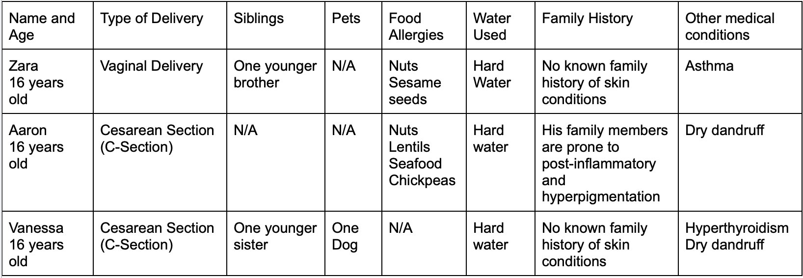
Survey Participants General Health Data
Student Reported Experiences with Atopic Dermatitis
Participant 1
Zara reported eczema flare-ups on her hands, face and the elbow area. Her thirteen year old brother does not have any specific skin conditions and he also had vaginal delivery. Normally, dry and cold conditions often impact the severity of her flare-ups. This suggests that the dry winters of Calgary may potentially impact the ability of Zara's skin to maintain moisture thus resulting in increased irritation. In comparison, Zara has also noticed increased irritation due to higher temperatures.
Zara noticed improvement in her eczema when she visited humid countries such as Malaysia and Indonesia. In Mexico, Zara's skin was more sensitive due to higher temperatures. When she visited Malaysia and Indonesia, the cloud cover was greater and the humidity was higher. This positively impacted her skin. She noticed no particular impact of hard water on her skin due to frequent use since early childhood. She also didn't notice any particular impacts of water on her skin when she travelled abroad.
Currently, Zara's eczema has significantly improved over time. Her experiences were more severe at 12 years old when she had moderate eczema. She describes her flares as irritating rather than painful. She rated her eczema at 12 years old a 4/10 on a severity scale.
Her allergic reactions to nuts and seeds include increased inflammation, itchiness, dryness and hives. She didn't notice effects of Atopic Dermatitis on her sleep nor any influence of sleep on her symptoms. She also didn't notice any significant changes due to air qualities in different countries.
Participant 2
Aaron reported flares on his forearm, arms and hands. Currently, he rates his eczema from mild to moderate. He reported that his eczema flares have worsened since his early childhood. He reported that dry and cold winter conditions cause irritation. He has visited humid and tropical regions such as the Southern state of Kerala in India. His eczema significantly improved in warmer and more humid environments. He reported that the monsoon or wet season of India helped improve his skin due to increased humidity. He noticed no particular impact of hard water on the skin but hot water did increase dryness.
His allergic reactions to nuts, lentils, chickpeas, and seafood include itchiness and anaphylaxis. He didn't notice effects of Atopic Dermatitis on sleep nor any influence of sleep on his symptoms. He also didn't notice any significant changes due to air qualities in different countries.
Participant 3
Vanessa reported eczema flare-ups on her hands and back. Her nine year old sister does not have any major skin conditions and she was also born through C-section. She described her eczema as very severe around age 8 (8/10), but currently rates it 2/10. She reported that dry environmental conditions increased irritation and redness. On her Florida trip, the high humidity significantly improved her rashes. Vanessa's severe eczema used to disrupt her ability to sleep. She described a procedure in which she would spread moisturizer and put on gloves to prevent moisture loss and to ease irritation. While Vanessa does not have any food allergies, the over-consumption of red meat increased irritation. She also reported that interacting with her dog initiated mild irritation rather than a severe reaction. She noticed no particular impact of hard water due to frequent use. She also didn't notice any significant changes due to air qualities in different countries.
Analysis
In general, all three participants reported that dry and cold conditions often increase irritation and itching. It is important to note that every person's eczema reacts in different ways to the environmental conditions. For instance, my eczema becomes increasingly worse in extremely high temperatures and locations with high humidity. However, the participants found that high humidity significantly improved their symptoms. Also, they did not feel any impacts because of hard water. However, my observations in Mangalore may suggest that hard water impacted my symptoms. Both Zara and Aaron have multiple food allergies which may suggest IgE mediated sensitivity. Finally, the type of delivery could have contributed to the severity of their eczema.
Conclusion
Conclusions
Internal health factors such as skin barrier dysfunction, immune sensitivity and gut health impact the susceptibility to Atopic Dermatitis from early childhood. External triggers like climate, humidity, allergens and water quality can further irritate and increase symptoms in Atopic individuals. Some lifestyle related factors which influence the severity of one's eczema include sleep, diet and the presence of food allergies. Overall, the dynamic interactions between these factors can increase bacterial infections and severity. To conclude, every individual with Atopic Dermatitis experiences these factors in a different ways. Skin barrier dysfunction and immune sensitivity is a unique and complex topic which varies from patient to patient. Therefore, the symptoms of eczema due to these interactions also vary.
Limitations & Further Study
Atopic Dermatitis and relevant topics about skin conditions involve a complex mix of genetics, internal factors and external triggers. Therefore, there is a lot of medically intense information which cannot be covered to a great extent in depth. There is still ongoing research on the different parts of the immune system and how they impact eczema. For instance, the role of T Regulatory Cells (Treg cells) in Atopic Dermatitis is still being studied due to complexity.
Citations
Agrawal, R., Wisniewski, J. A., & Woodfolk, J. A. (2011). The role of regulatory T cells in atopic dermatitis. Current Problems in Dermatology, 41, 112–124. https://doi.org/10.1159/000323305
Al‐Dhubaibi, M. S., Mohammed, G. F., Bahaj, S. S., AbdElneam, A. I., Al‐Dhubaibi, A. M., & Atef, L. M. (2025). The role of keratinocytes in skin health and disease. Dermatological Reviews, 6(2). https://doi.org/10.1002/der2.70028
Calgary Air Quality Index (AQI) : Real-Time Air Pollution. (2026b, February 28). AQI.in. https://www.aqi.in/ca/dashboard/canada/alberta/calgary
Čelakovská, J., & Bukač, J. (2016). Eosinophils in patients suffering from atopic dermatitis and the relation to the occurrence of food allergy and other atopic diseases. Food and Agricultural Immunology, 27(5), 700–710. https://doi.org/10.1080/09540105.2016.1148669
Climate & weather averages in Calgary, Alberta, Canada. (n.d.). https://www.timeanddate.com/weather/canada/calgary/climate
Climate & weather averages in Mangaluru, Karnataka, India. (n.d.). https://www.timeanddate.com/weather/india/mangalore/climate
Crna, R. N. M. (2024, January 9). Tips for Sleeping Better When Living with Severe Eczema. Healthline. https://www.healthline.com/health/atopic-dermatitis/sleeping-severe-eczema#eczema-at-night
DermNet. (2023, March 28). Barrier function in atopic dermatitis. DermNet®. https://dermnetnz.org/topics/barrier-function-in-atopic-dermatitis
DermNet. (2023a, March 22). Causes of atopic dermatitis. DermNet®. https://dermnetnz.org/topics/causes-of-atopic-dermatitis
Eczema Facts. (n.d.). National Eczema Association. https://nationaleczema.org/eczema-facts/
Eczema Society of Canada. (2022, May). Five tips for summer eczema. Eczema Society of Canada. https://eczemahelp.ca/five-tips-for-summer-eczema/
Eczema Society of Canada. (2026, January 15). Understanding the neuroimmune link in atopic dermatitis. https://eczemahelp.ca/understanding-the-neuroimmune-link-in-atopic-dermatitis/
Engebretsen, K., Johansen, J., Kezic, S., Linneberg, A., & Thyssen, J. (2015). The effect of environmental humidity and temperature on skin barrier function and dermatitis. Journal of the European Academy of Dermatology and Venereology, 30(2), 223–249. https://doi.org/10.1111/jdv.13301
ESCAdmin. (2024b, November 1). What is the role of the skin barrier in people with atopic dermatitis? Eczema Society of Canada. https://eczemahelp.ca/what-is-the-role-of-the-skin-barrier-in-people-with-atopic-dermatitis/
Eczema physical and Emotional effects | Rady Children’s Health. (n.d.). RCH. https://www.rchsd.org/programs-services/dermatology/eczema-and-inflammatory-skin-disease-center/physical-and-emotional-effects/
Fld. (2025, February 26). Understanding the layers of your skin. Florida Dermatology and Skin Cancer Center. https://fldscc.com/understanding-the-layers-of-your-skin/
Impact of water on atopic eczema prone skin. (n.d.). BIODERMA | Official Website Canada. https://www.bioderma.ca/en/your-skin/very-dry-sensitive-skin-prone-atopy/impact-water-atopic-eczema-prone-skin
Inquiry into biology.(2007).Mcgraw-Hill Ryerson. (PHYSICAL TEXTBOOK)
Institute for Quality and Efficiency in Health Care (IQWiG). (2025, April 22). In brief: How does skin work? InformedHealth.org - NCBI Bookshelf. https://www.ncbi.nlm.nih.gov/books/NBK279255/
IQAir. (n.d.). Calgary Air Quality Index (AQI) and Canada Air Pollution | IQAiR. https://www.iqair.com/ca/canada/alberta/calgary
IQAir. (n.d.-b). Mangalore Air Quality Index (AQI) and India Air Pollution | IQAIR. https://www.iqair.com/ca/india/karnataka/mangalore
Khosravi, A., Glińska, J., & Barańska-Rybak, W. (2024). Sleep efficiency and neurocognitive Decline in atopic dermatitis: a Systematic review. Acta Dermato Venereologica, 104, adv40459. https://doi.org/10.2340/actadv.v104.40459
Kim, B. E., & Leung, D. Y. (2018). Significance of skin barrier dysfunction in atopic dermatitis. Allergy Asthma and Immunology Research, 10(3), 207. https://doi.org/10.4168/aair.2018.10.3.207
Kim, J. E., & Kim, H. S. (2019). Microbiome of the skin and gut in Atopic dermatitis (AD): Understanding the pathophysiology and finding novel management strategies. Journal of Clinical Medicine, 8(4), 444. https://doi.org/10.3390/jcm8040444
Lopez, D. J., Singh, A., Waidyatillake, N. T., Su, J. C., Bui, D. S., Dharmage, S. C., Lodge, C. J., & Lowe, A. J. (2022). The association between domestic hard water and eczema in adults from the UK Biobank cohort study. British Journal of Dermatology, 187(5), 704–712. https://doi.org/10.1111/bjd.21771
Luschkova, D., Zeiser, K., Ludwig, A., & Traidl-Hoffmann, C. (2021). Atopic eczema is an environmental disease. Allergologie Select, 5(01), 244–250. https://doi.org/10.5414/alx02258e
Mangalore Air Quality Index (AQI) : Real-Time Air Pollution. (2026, March 1). AQI.in. https://www.aqi.in/in/dashboard/india/karnataka/mangalore
Murrell, J. (2023, December 7). What lipids do for your skin. Walter John Murrell, M.D. https://wjohnwmurrell.com/what-lipids-do-for-your-skin/
Professional, C. C. M. (2026, January 14). Dermis. Cleveland Clinic. https://my.clevelandclinic.org/health/body/22357-dermis
Professional, C. C. M. (2026b, February 10). Eosinophils. Cleveland Clinic. https://my.clevelandclinic.org/health/body/23402-eosinophils
Professional, C. C. M. (2025, December 29). Hypodermis (Subcutaneous tissue). Cleveland Clinic. https://my.clevelandclinic.org/health/body/21902-hypodermis-subcutaneous-tissue
Professional, C. C. M. (2026d, February 16). Immunoglobulin E (IGE). Cleveland Clinic. https://my.clevelandclinic.org/health/body/ige
Professional, C. C. M. (2026b, February 10). Keratin. Cleveland Clinic. https://my.clevelandclinic.org/health/body/23204-keratin
Quaresma, J. a. S. (2019b). Organization of the skin immune system and compartmentalized immune responses in infectious diseases. Clinical Microbiology Reviews, 32(4). https://doi.org/10.1128/cmr.00034-18
Research, G. P. F. E. (2025, June 26). Eczema types — Global Parents for Eczema Research. Global Parents for Eczema Research. https://www.gper.org/blog/types
Radhakrishnan, J., Kennedy, B. E., Noftall, E. B., Giacomantonio, C. A., & Rupasinghe, H. P. V. (2024). Recent Advances in Phytochemical-Based Topical Applications for the Management of Eczema: A Review. International Journal of Molecular Sciences, 25(10), 5375. https://doi.org/10.3390/ijms25105375
Rios-Carlos, M., Cervantes-García, D., Córdova-Dávalos, L. E., Bermúdez-Humarán, L. G., & Salinas, E. (2024). Unraveling the gut-skin axis in atopic dermatitis: exploiting insights for therapeutic strategies. Gut Microbes, 16(1), 2430420. https://doi.org/10.1080/19490976.2024.2430420
Russo, F., Zink, A., Magnolo, N., Scala, E., & Scala, E. (2025). Atopic Dermatitis and climate: Environmental stressors and care strategies. Dermatology and Therapy, 15(12), 3479–3493. https://doi.org/10.1007/s13555-025-01560-6
Silverberg, J. I., Hanifin, J., & Simpson, E. L. (2013). Climatic factors are associated with childhood eczema prevalence in the United States. Journal of Investigative Dermatology, 133(7), 1752–1759. https://doi.org/10.1038/jid.2013.19
T cells protect against COVID-19 in absence of antibody response. (2026, February 11). National Institutes of Health (NIH). https://www.nih.gov/news-events/nih-research-matters/t-cells-protect-against-covid-19-absence-antibody-response
UCF Health. (2022, May 6). Eczema vs. Atopic dermatitis: Causes\, Diagnosis\, treatment | Orlando | UCF Health. https://ucfhealth.com/our-services/dermatology/eczema-vs-atopic-dermatitis/
Vaneckova, J., & Bukač, J. (2016). The severity of atopic dermatitis and the relation to the level of total IgE, onset of atopic dermatitis and family history about atopy. Food and Agricultural Immunology, 27(5), 734–741. https://doi.org/10.1080/09540105.2016.1183598
Acknowledgement
I would like to acknowledge my parents who supported my idea of researching the several factors which impact Atopic Dermatitis. I would also like to thank Mr.MacLean for clearing my doubts on specific ways to research my data and improve my project. Lastly, I would like to thank my classmates for providing their personal insights and experiences with Atopic Dermatitis. Their contribution allowed me to explore the complexity of eczema.