Out the 3 most commonly used gene-editing technologies, which one is ideal for gene therapy in somatic cells via homology directed repair?
Yasmita Bhandari
Calgary Girls Charter School
Grade 7
Presentation
No video provided
Problem
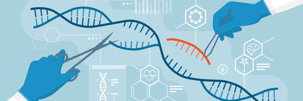
My question is “out of the 3 most commonly used gene-editing technologies, which one is ideal for gene therapy in somatic cells via homology-directed repair?” What I am exactly trying to explain is that from the 3 most commonly used gene editing technologies -CRISPR-CAS9, TALEN’s and ZFN- which one would be best when we did it in specifically somatic cells that make up parts such as tissues and that would not pass on these mutations to future offspring like germline cells would and we used Gene knock-in such as HDR?
Method
Method
- What is DNA?
- What is RNA?
- What is gene-editing?
- What is gene-therapy?
- What is the cell?
- What are organelles?
- What is the nucleus?
- What shape is DNA stored in?
- What are chromosomes?
- What are prokaryotes?
- What are eukaryotes?
- What is the difference between prokaryotes and eukaryotes?
- What are endonucleus?
- What is CRISPR-cas9?
- How does CRISPR work in a bacteria's immune system?
- What is a guide RNA?
- What is a PAM sequence?
- How does CRISPR-cas9 work in the body?
- What are the cas9 domains?
- When was CRISPR-cas9 invented?
- How is CRISPR-cas made in the lab?
- What is protein synthesis?
- How does protein synthesis happen?
- What are proteins?
- How does transcription work?
- How does translation work?
- What is ZFN?
- How was ZFN discovered?
- When was ZFN invented?
- How does ZFN work?
- What was the protein in ZFN?
- How is ZFN made in the lab?
- When was TALEN discovered?
- How was TALEN invented?
- How does TALEN work?
- What is NHEJ?
- How does NHEJ work?
- What is HDR?
- How does HDR work?
- How is CRISPR-cas 9 inserted in humans?
- How is ZFN inserted in humans?
- How is TALEN’s inserted in humans?
- What are the pros and cons of CRISPR-cas9?
- What are the pros and cons of ZFN?
- What are the pros and cons of TALEN’s?
Research
MOLECULAR STRUCTURES: Basic Living Unit Do you know the name for all living things? If not, they are called organisms. Organisms are living individual animals, plants or single-called bacteria. All living organisms are made up of something called the cell. Cells are the smallest unit that can carry out life. A cell's purpose is to provide structure for the body and convert nutrients within the body into energy, etc. Cells work together in groups and those groups are called tissues. After which the tissues gather up and work together to create organs. A thing to note would be that not all cells have the exact same purpose and function. As stated earlier, cells are tissues and tissues are organs. So that would mean that there would be different functions of cells depending on the organ. Another intriguing fact about cells is that they are produced from other cells. The process of new cells being made is called mitosis and meiosis. In this process, one cell splits itself into two cells, and so on, causing a continuous stream of cells or one cell splitting into two. This also means that all of us started as a single cell and divided until there were trillions of them, forming us. Although the cell carries out so many functions and keeps us alive, they are astonishingly minuscular. Most cells are no bigger than 0.1 to 100 micrometers in diameter. The cell also has organs inside of it that do the jobs required. They are called organelles. Think of it as the organs in the human body but at a minuscule size. Although there are a great deal of organelles within an animal cell, 13 of them are major. Just to name a few would be the mitochondria -used to break down sugar molecules to convert into energy- , ribosomes -significantly important for protein synthesis- and the nucleus. The nucleus is an exorbitantly important organelle. The nucleus carries something called chromosomes, which are vast, compact strands of material which contain a human’s genetic blueprint, the DNA. There are also two types of cells classified in gene-editing. Those two cells are called somatic cells and germline cells. S Somatic cells are diploid cells, which means they contain pairs of chromosomes. Any mutations that occur in somatic cells only affect the individual that has the cells and will not affect the offspring or future generation. Germline cells are cells that do affect the offspring and future generations. Examples of germline cells are reproductive parts such as sperm and eggs. Germline cells are haploid, meaning there is one chromosome in the cell. This is because when reproducing, both chromosomes from the mother and father chromosomes will be present. DEOXY-RIBOSE DNA, located in the nucleus of the cell, is a molecular structure that withholds every gene within an organism. DNA has the instructions for protein synthesis which is mandatory for the growth of an organism. The DNA is within the nucleus and is held in the structure of chromatin. DNA stands for Deoxy-RiboNucleic Acid. The name directly correlates with the structure of it. DNA is primarily known for its double-helix structure. Due to this, DNA looks like a twisted ladder. DNA is held together by nitrogenous bases and hydrogen bonds between the bases that hold the bases together. Although hydrogen bonds are weak, they do hold the bases due to the many amounts of the bonds between bases. DNA has 4 nitrogenous bases that pair together and are complementary. The 4 bases are adenine, thymine, cytosine, and guanine. Adenine and thymine pair with 2 hydrogen bonds between them whilst cytosine and guanine pair with 3 hydrogen bonds between them. The bases in DNA are classified as Purines and Pyrimidines. These 2 structures fit perfectly with each other. The Purines in DNA would be adenine and guanine while the pyrimidines would be cytosine and thymine. The DNA has a back bone that is made of alternating 5-carbon sugar- deoxyribos- and phosphate. These nucleotides within a singular strand of DNA are compactly held together by insanely powerful bonds where two atoms share one or more pairs of electrons called covalent bonds. These kinds of bonds are stronger than hydrogen bonds. Now let’s talk about DNA from a wider perspective. We already have stated that DNA is held within the nucleus, but in what structure. The DNA is compactly wrapped around proteins called histones. Afterwards, they wrap into something called a chromosome. The human body contains 46 chromosomes and 23 pairs. 22 of them being autosomal chromosomes and the twenty-third one being a sex chromosome. RIBO-NUCLEIC ACID RNA is a crucial and imperative structure in molecular biology. It is a versatile structure that accomplishes a myriad of tasks in the cell from carrying out genetic material of the DNA from the nucleus and enforcing it to become proteins to regulating genes and acting as enzymes, RNA will do it all. RNA is found in various places within the cell. These include inside of the nucleus and in the cytoplasm. RNA’s full form is ribonucleic acid, similar to DNA’s full form. That it isn't a coincidence. RNA and DNA are chemically analogous whilst also having noticeable differences. If you look at the full form of these terms -deoxyribonucleic acid and ribonucleic acid-, you can notice the similarities. Both of them sound identical except that DNA has the term “deoxy” added towards the front. That is because DNA’s backbone consists of a singular less oxygen molecule than RNA, thus the term “de-oxy-ribose”. This also means that RNA’s backbone is made up of phosphate and ribose sugar. This is because ribose is a little less stable and more flexible, fitting the role of rna perfectly. There is also another major difference between these two. The difference being that RNA is single-stranded, and not double-stranded like DNA. This is a transparent difference between DNA and RNA. Let’s talk about the similarities of aforementioned terms. DNA and RNA are composed of identical chemicals. RNA, like DNA, has nitrogenous bases. RNA has adenine, guanine and cytosine but it lacks thymine. Alternatively, it has a chemical more suited towards its purpose, uracil. RNA is short-lived compared to DNA, and uracil is less energy consuming compared to thymine, thus it is used instead. RNA is truly captivating with its complexness and important to understand. Prokaryotes and Eukaryotes Prokaryotic and eukaryotic cells are a major factor in our world. Although their names may sound similar, they do have noticeable differences. Let’s first proceed by talking about prokaryotes. Prokaryotes are single celled organisms that primarily consist of a simple skeleton and lack many organelles that are found within a eukaryotic cell. They lack important organelles membrane-bound organelles such as mitochondria and the nucleus. Instead of the nucleus, they have a nucleoid that floats around in the cytoplasm of the prokaryote. Prokaryotes rule over the domain of archaea and bacteria, with approximately 50% percent falling in that category. Now let’s talk about eukaryotes. As you may have assumed by now, eukaryotic cells are more complex than prokaryotes. And they also make up more complex structures. Eukaryotic cells have more organelles such as the nucleus, mitochondria and much more. Eukaryotic cells make up many living organisms such as animals, plants, fungi and seaweed! Both of these are crucial for the world and environment. And there is a fundamental part in prokaryotes that makes a gene-editing tool succeed.
CRISPR-CAS9: History of CRISPR CRISPR-cas9 is a ground-breaking discovery for all the science fields in the world, but do we know of the brilliant minds who made it come true? They were Nobel prize in chemistry winner, microbiology professor at the University of California, Jennifer Anne Doudna and Nobel prize in chemistry winner, founder of the max planck program for the science of pathogens, Emmanuelle Charpentier. Both of them made history for this is the first time that the Nobel prize has been awarded to 2 women. It is said that they are glad it was received by 2 women to empower and drive the younger generation of girls and women. But crisper wasn’t an overnight miracle, it was intensive research and hard-work. And it wasn’t also just discovered by 2 women, it started earlier than that, with a lot more hands helping. The first observation and discovery of CRISPR happened in 1987, at Osaka university, Japan. The appearance of CRISPR was spotted by Ishino Et Al within Escherichia coli bacteria. Ishino spotted this when looking at the DNA sequence of the bacterium and seeing the CRISPR pattern. The sequencing of these DNA fragments took many months of time, but even so, the potential and origin of it failed to be deeply understood by its discoverers. But there were proposals of how this could be used to egenotype -process of determining the DNA sequence- many strains of bacteria. Another huge leap for the journey happened in 2005, at the university of Alicante, Spain, where it was determined that CRISPR was an adaptive immunity system in bacteria. Afterwards, there were more break-through for the CRISPR technology such as detecting that CRISPR is an adaptive immunity but not as big as when 2 scientists combined their knowledge to create a working tool. In 2011, Emmanuelle Charpentier discovered something fascinating while researching on a bacteria -streptococcus pyogenes-, tracrRNA. TracrRNA is a factor in making guide RNA for the CRISPR protein, thus making itself a crucial ingredient for the creation of CRISPR. While Charpentier was busy investigating her new discovery, Jennifer Doudna was mapping -variety of ways to determine relative positions of markers- CRISPR proteins. Luckily, these two ideas would soon combine. In spring of 2011, both Charpentier and Doudna attended an academic conference about CRISPR. This led to their first meeting. Both of them decided to go to a cafe and talk about their discoveries and information on CRISPR, making a new path open up. After the meet up, both scientists started actively collaborating on CRISPR. Which ultimately led to it being conjured in 2012, a giant leap for mankind. Bacteria’s Immune System Before going into CRISPR, we need to learn about something that actually makes it successful. To learn, we need to first understand a bacteria’s immune system. Surprisingly, CRISPR-Cas9 is a natural process that happens inside a prokaryotic cell bacteria and archaea. It works as an adaptive immunity system to protect the single celled organism from bacteriophages: a type of virus that can infect bacteria and archaea. Natural Cas9 works in 2 segments. The first segment is something called clustered regularly interspaced short palindromic repeats, also known as CRISPR. The bacteria was a separate region dedicated for storing crisps called the bacterium genome. And the second segment being Cas9 enzymes. The Cas9 is a nuclease: an enzyme that binds and can create double stranded breaks in DNA sequence. Now let’s talk about the process when a phage actually enters the bacterium. Bacteriophages are shaped like minuscule moon launchers, filled with viral DNA inside of it. The phage is attached to the surface of the bacterium and releases it within the bacteria. Once inside, it can cause havoc. This is where two enzymes called cas1 and cas 2 begin their duty. Both of these are different enzymes but they are joined together partially in this instance. These enzymes scan through the viral DNA, searching for a piece of DNA to cut. But they don’t just randomly cut the DNA wherever they wish. They have to find a specific PAM -protoscape adjacent motif-. In this instance, the motif is any nitrogenous base followed by two guanine bases. After the enzymes detect them, they make a precise cut upstream of the PAM. The segment of cut DNA then goes on to be called protospacer. This fragment then gets stored in a part of the bacterium genome, called the CRISPR array. CRISPR is the part of the bacteria’s genome where all of these segments of viral DNA are kept. It follows the pattern of a protospacer and repeats. After the protospacer is cut, it is inserted in the 5’ primer end of the CRISPR region. After being inserted, a repeat is formed. The protospacer then becomes a spacer. Every repeat in the crispr array is identical. The crispr array is also very flexible. What I mean by this is that it can have a myriad of spacers and repeats or barely just one, depending on how many times the bacteria has been infected. This entire process also applies to the full form of CRISPR as well. Clustered Regularly Interspaced Short Palindromic Repeat. Occasionally, a RNA polymerase will transcribe -making a RNA copy of DNA- the entire CRISPR array. This strand of RNA is called pre-crRNA. The strand has the spacers and repeats as well as a new addition called unprocessed tracrRNA. TracrRNA is a part of something called “crispr locus” in the vicinity of the crispr array. These segments are also transcribed by the RNA polymerase. The tracrRNA also consists of regions that are complementary to some regions of crRNA. This causes those complementary parts to attach and bind. Afterwards, an enzyme called RNAase goes through the strand and starts cutting segments. The cut parts will consist of the RNA of the spacer, the repeat and the tracrRNA. This RNA molecule sequence is called car:tracrRNA. This molecule then gets picked up and attached to an enzyme called Cas9. Let’s discuss what would happen if a type of bacteriophage that has already infected the bacteria once were to enter once more. If that were to happen, the Cas9 enzyme that has the repeat withholding the information of the page would go scan the viral DNA released from it. Once it finds the PAM -any nitrogenous base followed by two guanine’s- , it will latch onto it and unwind the double helix structure. Afterwards, it will see if the guide RNA is complementary to the strand opposite of where the PAM sequence was found. If it is complementary, the Cas9 enzyme will use its domains and create a double-stranded break(DSB). It will continue the process throughout the viral DNA until it is not a threat. Now scientists observed this mechanism within a bacterium and pondered whether this could make a difference in the real world, leading to the ground breaking discovery of CRISPR. Cas9 Before continuing to talk more about crispr, let's talk about cas9. The key factor about cas9 that should be known is that it is the endonuclease responsible for doing the DSB(double-stranded break). But cas9 itself is a pretty complex enzyme on its own. Let’s just talk briefly about it and its parts. The cas9 enzyme itself is a long piece of poly-peptide consisting of six domains. Pi: Pam interacting domain. This domain does the job of identifying the correct PAM, thus the name Pam interacting domain. RuvC and HNC nucleases domains are the part of cas9 that actually cut the DNA. The other parts are called REC1, REC2 and bridge helix. These structures are typically there to hold the guide RNA in place. Now without further distractions, let's dive into CRISPR-cas9 working in the human genome. In a lab: CRISPR-Cas9 Now, we know about the gRNA and the tracrRNA, but how do they work in a human body? Well, when scientists were researching this, they found that they could join the two gRNA and the tracrRNA using something called a linker loop. This was very important because two different strands could not do gene-editing efficiently. After the strands combine, they are called sgRNA, an abbreviation for Single-Guide RNA. The RNA base then gets modified to fit the DNA sequence and is bonded to the Cas9 protein. CRISPR: In The Body Now that we have stated the basics and fundamentals, let's dive into how Cas9 performs in the human genome. We have already stated how Cas9 is made in the lab and the tools engraved in it lets drive straight into the mechanics. When Cas9 enters the genome domain, its first and most important goal is to find the correct gene that is causing the issue. This does so with the assistance of the gRNA (Guide RNA). Cas9 scans along the strands of DNA using its PAM interacting domain until it can detect a sequence that is the PAM sequence -Protospacer Adjacent Motif-. The PAM sequence to be found is once again, any nitrogenous base followed by two guanines The genomic target of Cas9 is a twenty nucleotide sequence. Cas9 then begins to unwind the DNA sequence. Cas9 uses something called ATP independent to follow through with the process. After Cas9 unwinds the dna, the RNA binds with the dna strand, forming a R ring. A R ring structure is the structure that the DNA and RNA form when bonded. If the guide RNA is not a sufficient match, then the cas9 enzyme will continue to move forward and find the correct match. As explained earlier, if it finds a PAM, it will then go to the opposite strand of DNA that is upstream of the PAM to see if it's complementary. If the RNA matches us correctly to the DNA sequence, the Cas9 uses binding energy -energy that is formed after weak interactions between complementary and substrates and enzymes- and its two nuclease cutting domains -RuvC and HNC- to make a double stranded break on the DNA. This cut will often occur approximately four nitrogenous bases upstream the Pam. The reason I say approximately is because it can also be three or five bases upstream. But it does typically fall within the 3-5 stamp. Cas9’s purpose then gets fulfilled and the gene is cut. Gene-cutting isn't the only thing Cas9 is capable of performing. In some cases, scientists may disable one or more of the cutting domains of Cas9 and attach another enzyme to it. This gives Cas9 the role of a carrier. For example, a deaminase -an enzyme capable of mutating a specific nucleotide- can be attached onto Cas9. Cas9 will then go to the sequence instructed by the guide RNA. And rather than cutting, it will unravel the DNA and allow the deaminase to mutate the gene. This could make a gene that does not belong make sense, such as perhaps turning adenine to guanine, if it is in the strong segment, causing a mutation.
TALEN’s: History of TALEN’s The history and creation of TALEN really began in 1989 when the TALE protein was discovered in xanthomanous oryzae. But it was not yet discovered or thought that it could be used as a gene-editing tool. In 2007, when a more thorough erase was happening on the TALE protein, a researcher scientist working in the Ulla Bonas group in the Martin Luther university, Halle, Wittenberg saw a innovation, he and his colleague were trouble shooting a problem where two different TALE proteins were activating the same gene. That's when he came to a realization and basically said “why not look at it as if one of those RVDs recognized one base”. He and his colleague wrote down this pattern of how the 12th and 13th amino acids were different from the others At the same time, plant geneticist Adam Bogdanove and his graduate student Matthew Moscou came to the same conclusion. Both of these groups published their own research in October of 2009. And then in the year of 2010, the TALEN method was developed by many scientists including the ones listed above. Xanthomonas Oryzae Now let's talk about where TALEN’s are found/ Talens naturally occur in a plant pathogenic bacteria called Xanthomonas oryzae. This bacteria typically infects plant life and results in white or yellow splotches on it. This bacteria enters the plant's system via its stomata and infects the plant cells. The Xanthomas Oryzae then releases a protein called TAL -transcription activator-like- effectors. This protein then proceeds to infect the plant. But it only infects specific genes. Before talking about the genes it can and cannot infect A plant cell contains two types of genes. One of them being R genes (resistant genes). R genes are genes in a plant's genome that are meant to revisit and protect the plant via the creation of pathogens. These proteins detect specific substances called effectors that are produced by a pathogen. A fact about R genes is that they are dominant genes -genes that most likely occur when organisms reproduce- and are only sometimes recessive. The other type of gene are called S genes (susceptibility genes) and are negative regulators in plants. These genes are hosts and can be easily exploited by pathogens. This can lead to various different issues for the gene can increase the bacteria’s growth and plays a critical role in disease development. Knowing this, let's go back and talk about the TAL effector entering the plant cell. After entering the plant cell, it heads straight to the nucleus. There, it binds to the S gene. After binding, it increases that specific protein’s production of protein synthesis. This is why it gets its name “transcription activator like” effector because it increases the transcription of the protein. This helps the bacteria grow and develop. Now, a question arose. We already know that the TAL effector only binds to S genes, but how does it know to recognize one? Scientists naturally fuelled by curiosity decided to study the TAL effector. In the protein, they found a N-terminal or also known as A/B terminal at the beginning of the protein. A N-terminal in a protein is a free amino group consisting of -NH2, thus giving its name of N-terminal. This protein also has a c-terminal at the end of it that is made up of a free carboxyl group. The effector also has an acidic domain that increases transcription and a NLS -nucleus localization signals- domain that guides it to the nucleus. The protein also has a crucial region for recognition called the central repeat domain. The domain has 1.5 to 33.5 repeats and each repeat may recognize anywhere from 2 to 34 DNA bases. Each repeat also consists of 33 to 35 amino acids that actually recognize the DNA. When scientists started to study it carefully, they noticed a pattern. All the amino acids were the same and repeating with the sole exception of the 12 and 13th amino acids. They saw that repeatedly in every unit to the TAL effector, the 12 and 13 amino acids were different and varied very repeatedly. This led them to realize that it was the variation of the 2 amino acids that was specific for the recognition. These amino acids are called repeat variable diresidue or simply, RVD. Each amino acid pattern determines the base that they will bind to. For example, if the amino acid is HD, then it will bind to cytosine. Or if the amino acid is NI, then it will bind to adenine. When it comes to the actual binding, the tal effector searches the nucleus until it finds the strand that corral the to its amino acids and binds to it. When scientists saw this, they wondered whether they could harness this technique and use it on humans. In The Cell: TALEN’s After being engineered in the lab and bonded to a DNA binding domain, a is complete. This can either be transferred into a cell via ex-vivo or in-vivo. After entering the cell, it goes to the nucleus. In the nucleus, it starts to go through all of the DNA sequence. Once the TALE’s find the correct recognition sites using the Amin acids in them, the Fok1 protein binds to it. The Fok1 also must find a recognition sequence that is 5’-GGATG-3’ and 3’-CCTAC-5’. Since the protein is a dimmer protein, it must wait for the other half to arrive. Once the 2nd dimmer arrives and binds to the sequence on the complementary strand, these two proteins bind to form a polymer. This results in a double stranded break 13 and 9 nucleotides downstream the complementary stand. This break has an almost diagonal type of structure to it.
ZFN: History of ZFN Let's talk briefly about the history of ZFN and how they were discovered and the process through all of it. There are two major components that were needed to be discovered for ZFN to work. The first major component was discovered in 1981. This component was an endonuclease called fok1. Fok1 was discovered in a bacteria called flavobacterium okeaokoites. This was a major discovery for the endonuclease district The second major step was in 1985 when a group of scientists led by Aaron Klug were studying the genome of an African clawed frog called Xenopus Laevis. While studying, the scientists observed something unusual. They saw that the rna polymerase wasn’t eating a certain stand, and when they researched even further, they discovered the zinc finger motif, which as you may supposedly tell is vital for the gene-editing technology. In 1996, zinc finger nucleus genetic engineering was successfully discovered by Srinivasan Chandrasegan and his team. They conjured this technique by fusing an engineered dna binding domain and a non-specific cleavage domain, Fok1. Protein Synthesis I know that you’ve heard that DNA is the blueprint to your body or that DNA is the guide for making proteins. But what does that really mean? What they are most likely talking about or implementing is protein synthesis. Our genes have the code for the traits of our body but for them to be implemented, proteins need to be made. Protein synthesis is the process of our DNA information being turned into proteins. Proteins are unimaginably important for they make structures for our body, act as enzymes, resulting in our traits and much more! Let’s talk very briefly about it. Protein synthesis includes two major steps to make it succeed, called transcription and translation. The first step is transcription and it occurs in the nucleus. At the beginning of the process, an enzyme called RNA polymerase with the assistance of proteins called transcription factors identifies and latches onto a region of the DNA strand where a gene begins called a promoter. After binding with the promoter, the RNA polymerase pries it open. The strand that is 3’ to 5’ is used as the template or also known as the antisense strand. This strand is the strand used to create mRNA or messenger RNA. The opposite strand is called the non-template strand or the sense strand. The RNA polymerase then continues to move along and reads the nitrogenous bases. While doing this, the RNA polymerase takes free RNA bases in the nucleus that are complementary to the antisense strand and forms a mRNA. The mRNA is then formed in a 5’ to 3’ direction. This process of the RNA polymerase moving along the strand is called elongation. The RNA polymerase also fixes the DNA back to its original form while working, leaving only ten to twenty bases exposed. After the RNA polymerase reaches the end of a gene, a process called termination occurs. This is when the RNA polymerase detaches from the DNA strand. But at the end of transcription we have mRNA. MRNA carries the genetic information of the gene copied and escorts it out of the nucleus to turn into proteins. But prior to that happening, the mRNA goes under mRNA processing. This is when some modification occurs to the mRNA to make it ideal for translation. In this process the mRNA gets checked and some parts may get deleted and etc. After the modification is done, the mRNA proceeds to exit the nucleus. This is where translation begins. After leaving the nucleus, the mRNA proceeds to make its way through the cytoplasm to a ribosome. As explained earlier, a ribosome is what turns the mRNA’s information into proteins. A notable factor of ribosomes would be that they are made up of rRna’s. The mRNA strand has many bases but every three bases are what code for the protein. Every three bases in the mRNA is called a codon. This is important due something called tRNA’s. TRNA’s have amino acids and these are important for translation. A small ribosomal sub unit binds to the mRNA and brings in something called an initiator tRNA. Then the large ribosomal sub unit joins to complete the complex. Let’s talk a bit about tRNA, also known as transfer RNA has a structure where there are three nitrogenous bases that are complementary to the given mRNA strand. Every tRNA also has an amino acid attached to it. In total, there are approximately two to fifty amino acids used in doing protein synthesis.. As the ribosome reads the mRNA strand, the complementary tRNA binds to it, leaving behind the amino acid attached to it. The start amino acid is typically methionine and the ending is typically a C-terminal made up of an unbound carboxyl group. We can tell which amino acids will be given via using a codon chart. After the mRNA strand is read and the amino acids are submitted, a long chain of amino acids is formed, called a peptide chain. This structure will then form itself into a complex protein shape, thus applying its function in our body. The protein that is made also goes through many forms and structures. The first form it goes through is called the primary protein structure. This is the original structure that is made. Afterwards, the bonds in the amino acids form secondary structures. These structures are beta sheaths and alpha helix. These proteins then combine and form a tertiary protein structure. Once two tertiary protein structures combine, they form a quaternary protein structure. Xenopus Laevis Now that we know the basics of zinc finger’s, the history of ZFN and different protein structure’s, we talk about what role it played in a frog's genome. Before proceeding, I have talked about the term “genome studying” before but haven’t fully given it a clear definition. The term genome studying means observing and seeing which genes affect what trait in the organism. This also means seeing how protein synthesis works in the genome. So when scientists were studying the genome of the Xenopus Laevis, they observed something abnormal in the protein synthesis process. You see, while other gene segments were being transcribed how they are normally supposed to, certain genes were not being transcribed. These genes were the genes that were producing 5s ribosomal RNA that made up the ribosome. The issue was that the RNA polymerase was not initiating transcription, thus not transcribing. Then scientists observed something else. Some that opened up doors to many paths. They saw that there was a way that the rna polymerase would initiate, and that way was when a protein would bind to it. This protein factor's name was transcriptional factor III A(TFIIIA). In other words, for transcription to be initiated, transcriptional factor III A must be binding to the RNA polymerase enzyme. Now this does beg the question, why? Why does there need to be a protein bound to the enzyme to initiate protein synthesis? The reason is because it creates a meta stable complex with the 5S DNA and it also assists forming stable and competent transcription complexes. Now scientists decided to investigate even further and find what exactly this protein was. A scientist named Engelke was the one who decided to isolate the TFIIIA protein and extract it from the genome. When researching, they found a 30 amino residue structure. Scientists then decided to look deeper into these 30 amino residue structures and saw that and learned that the protein was composed of secondary protein structures. The motif they saw of two parallel beta sheaths and an alpha helical structure. You might be wondering how they are connected and held together properly? This is due to Zn Ion. There is a zinc ion in the structure that's purpose is to stabilize the beta sheaths and the helical sheets. It does this by binding to the four amino acids. In secondary proteins, there also are two conserved amino acids that are bonded on top of the antiparallel beta sheaths called cystine while the alpha helical structure has two histidine proteins bonded to it. This zinc ion stabilized the entire structure. We call this structure a zinc finger motif. Now you may be curious how this could possibly relate with gene-editing? Well this all comes back in a circle the the alpha helical structure. Without going into too much detail and just summarizing it, the alpha helical structure has DNA, a binding domain that can recognize DNA sequences. In The Lab-ZFN In the lab, ZFN is modified for the DNA sequence. The alpha helical structure that is responsible for recognition gets changed and prepared. There can be two ways for this. The first way is called OPEN(olimogerized pool engineering). This method generates zinc finger arrays that have a high affinity for the DNA strand and chooses it. They are chosen by looking at the zinc finger module that shows the different arrays that are tailored to the gene that the ZFN might be used for. Afterwards, these customized ZFN take approximately eight weeks to create. ZFN: In The Cell Once ZFN enters the nucleus, it starts scanning the bases. Once ZFN finds the sequence that is complementary to the sequence of the Fok1 and its alpha helix’s, the recognition domains bind to it. The Fok1 itself covers six bases, making both of the proteins take up twelve nucleic bases. One of the zinc finger proteins can recognize one codon, also equivalent to three nucleic bases. A limitation with ZFN is that the highest number of zinc fingers that can be used is only four. Meaning that one side can only cover twelve bases, causing 24 bases to be covered overall with the zinc fingers. Once the ZFN is secure and binds to the dna, both of the Fok1 proteins on both sides bind together in almost a diagonal angle, causing a DSB.
REPAIR PROCESS: NHEJ When a double stranded break occurs whether via a gene-editing method of naturally, there are many ways to rebuild it, including Non-Homologous End Joining (NHEJ). When a DSB occurs, proteins called KU’s bind around the end of both cut DNA leaving a bit of DNA exposed. The KU protein then recruits the DNA PK Catalytic subunit, also known as DNA-PKcs. DNA-PKcs then recruits the nucleus Artemis and those two get phosphated. Artemis then proceeds to cut any lone single bases from the end of the DSB. Then an enzyme called ligase IV acting with a complex XRCC4 AND XLF cernunnos legates the broken ending together. Meaning that it joins the two ends. The con to this method is that it is aeroprone and typically causes mutation or silences a gene. Scientists, due to this, may add a DNA template for the cell to build off of. HDR-Natural Other than NHEJ, there are other mediums to gene repair. A natural way in the human body is HDR. HDR, standing for homologous directed repair, is done with a sister DNA strand. Our body typically has two identical chromosomes called sister strands. When a DNA break occurs,the sister strand can be used as a template. The first step is SDSA, or full form being, synthesis dependent strand annealing. In this process, a recBCD protein binds to the 5’ on both of the stands on the broken gene. This protein then proceeds to cut and resection the 5’ until it reaches a chi site. A chi site is a strand of DNA that contains the nucleic base sequence of “GCTGGTGG”. Note that a chi site can be anywhere and can even be hundreds of bases away from the breakage point. After a chi site is found, the 3’ end on the strand closest to the sister goes down and binds to the beginning to it. Then, an enzyme called DNA polymerase goes along the sister strand and the broken bottom strand, giving the broken 3’ a a strand till it reaches a chi site. Now, the attraction of the broken strand and the newly sequenced strand is strong due to the complementary bases. This causes the broken and newly made strand to join together. Then, the other strand with the 5’ to 3’ gets a new strand made by DNA polymerase due to there being a template strand. The strand now gets ligated by ligand 4, meaning it makes a phosphate bond. HDR-SYNTHETIC After talking about how homologous directed repair works naturally in the body, let's better understand how it can be performed via gene-editing. A DNA template is sent with the endonuclease or whatever protein is being used to cut DNA. This has the template of the DNA sequence that should have been there in the strand rather than the mutation. We know to find the template because we know where the DSB has occurred. The template DNA fragment is in the center of two other strands of DNA. We call these strand homology arms. There is a left and a right homology arm. Homology arms, as said in the name, are homologous to the strand of DNA surrounding the DSB. The process begins with a recBCD resectioning the 5’ to a chi side. Afterwards, the 3’ near the template DNA gets pulled down into the template DNA creating a D form. DNA polymerase then creates a strand of DNA following that template. Afterwards, the newly made strand connects to its original strand. This new strand is now somewhat of a template for the 5’ to 3’ end strand and DNA polymerase proceeds to make the new strand according to that.
ENTERING THE CELL: Invivo and ex-vivo There are many ways to get the gene-editing therapy inside of the cell. They are classified into two groups: In-vivo and ex-vivo. In-Vivo is inside the cell and ex-vivo is outside edited. One way to insert the medicine is by injecting the medication via IV into a specific tissue. Another way is to use a virus that acts as a vehicle to transport the medicine. Although, there are many risks because the virus might infect.
REAL WORLD EXAMPLES: CRISPR-cas9 - Real World CRISPR-cas9 can be used for various purposes to cure cancer, cystic fibrosis and sickle-cell disease. These diseases have affected humans a lot and crispr was there to help. A real life example of this happened with a baby named KJ. At birth, KJ was diagnosed with a rare urea cycle disorder called CPS1. CPS1 is a metabolic disease where the body cannot process or excrete proteins that you take in your diet. This will cause the toxic ammonia to go into your blood and then build up in your brain. The only cure up till then for CPS1 was a liver transplant. But KJ’s case was a more severe case and needed to be acted upon quickly. Then, scientists had an idea, CRISPR -cas9. Prior to KJ being born, scientists had still been working on a way to use CRISPR as a therapy medium but just could not crack the code. But when KJ was born, his gene variant cracked the code. After studying it, the scientists made a drug specifically for him in six months. On February 25, 2025, in the Children's Hospital of Philadelphia, KJ received the infusion for the first time. The CRISPR medication was immediately absorbed by the cells in the liver after it entered the blood stream. Afterwards, it cut the gene in the DNA that was causing the disease. Scientists predict that he will need more doses for it to be an efficient therapy. Although, there is a hope that after more treatments, KJ can live with little to none medications. KJ was the first patient to get a personalized treatment and we hope for there to be many more opportunities for people needed. ZFN:Real World Zfn has many uses like any other gene-editing technology, but it also has a remarkable feat. Zfn has been used and successfully completed to alter the CCR5 gene in humans. This gene codes for the T-cells. Due to this gene being altered and modified, the CCR5 has created almost like a resistance to hiv, a horrible disease. This was a huge step for gene-editing and much more. TALEN’s: Real World Unlike CRISPR and ZFN, TALEN’s prominent examples are in the field of agriculture. TALEN’s has been used in rice to engineer resistance to disease, droughts, and much more. TALEN’s has a huge impact on gene-editing in the field of agriculture.
COMPARE AND CONTRAST: Pros and cons: CRISPR-Cas9 Pros: * Easy to construct * Simple structure * Efficient * Good for gene-knockout * Short amount of time needed for construction *Low cost Cons: * High frequency chance for off-target mutations * Not precise enough *May cause cells to lose their cancer fighting abilities
Pros and cons: TALEN’s Pros: * Extremely precise * Low off-target mutation rates * Targets one base at a time Cons: * large size * Costly * Complex structure * Long construction time
Pros and cons: ZFN Pros: * Precise * Targets a codon at a time Cons: * Causes cytotoxicity * Complex structure * Long build time
Data
A site and reference that directly contributed to my questions was the site https://www.jax.org/news-and-insights/jax-blog/2014/march/pros-and-cons-of-znfs-talens-and-crispr-cas. This site talked about all 3 of the gene-editing technologies, including pro’s and con’s, which was massive help towards my research.
Conclusion
Conclusion After conducting my research, my hypothesis was rejected. The correct answer to the ideal gene therapy when looking consecutively at the factors of precision, off-target mutations and efficiency with homologous directed repair, the answer is TALEN’s. TALEN’s are extremely precise, more than ZFN and CRISPR-cas9, cutting the strand where needed. Its risk of having off-target break is low with less than 1% mutation rates whilst CRISPR-cas9 can have an off-target rate as high as 77%. This also answers the question of precision. In a study conducted on how efficient the gene-editing techniques are, TALEN stimulated a more efficient homology directed repair. Another reason is also that CRISPR has a blunt cut whilst TALEN creates a staggered cut, which may stimulate HDR more efficiently. In conclusion, the ideal gene-therapy in somatic cells when using the three most commonly used gene-editing is TALEN’s when looking for precision, accuracy and HDR editing.
Citations
References: https://pmc.ncbi.nlm.nih.gov/articles/PMC8380213/ https://www.jax.org/news-and-insights/jax-blog/2014/march/pros-and-cons-of-znfs-talens-and-crispr-cas https://link.springer.com/article/10.1007/s00253-010-2798-6 https://insights.som.yale.edu/insights/is-crispr-worth-the-risk https://pmc.ncbi.nlm.nih.gov/articles/PMC4916124/ https://blog.addgene.org/crispr-101-homology-directed-repair https://www.urncst.com/index.php/urncst/article/download/680/458/12890 https://www.news-medical.net/life-sciences/CRISPR-The-End-for-Zinc-Fingers.aspx https://en.wikipedia.org/wiki/Xanthomonas_oryzae https://pmc.ncbi.nlm.nih.gov/articles/PMC3694601/ https://scholar.google.ca/scholar?q=zinc+ion+in+xenopus+laevis&hl=en&as_sdt=0&as_vis=1&oi=scholart https://www.thoughtco.com/protein-structure-373563 https://www.chop.edu/news/worlds-first-patient-treated-personalized-crispr-gene-editing-therapy-childrens-hospital https://www.nature.com/articles/d41586-025-03847-2 https://www.genome.gov/genetics-glossary/Somatic-Cells https://en.vectorbuilder.com/resources/faq/crispr-vs-talen.html https://www.genengnews.com/insights/analyzing-talen-vs-crispr/ https://www.researchgate.net/post/what_are_the_pros_and_cons_of_CRISPR_than_the_other_gene_editing_assay_like_ZFN_TALEN https://www.azolifesciences.com/article/What-is-Cell-Biology.aspx https://synapse.patsnap.com/article/crispr-vs-talen-vs-zfn-which-gene-editing-tool-should-you-use? https://www.abcam.com/en-us/knowledge-center/dna-and-rna/gene-therapy https://www.biocompare.com/Editorial-Articles/576583-How-Does-CRISPR-Compare-with-Other-Gene-Editing-Methods/ https://www.joelsartore.com/ani117-00002/ https://en.wikipedia.org/wiki/African_clawed_frog https://pmc.ncbi.nlm.nih.gov/articles/PMC27934/#B8 https://www.sciencedirect.com/topics/biochemistry-genetics-and-molecular-biology/foki https://academic.oup.com/nar/article/36/6/2073/2410352 https://www.ncbi.nlm.nih.gov/books/NBK609557/ https://en.wikipedia.org/wiki/FokI https://pmc.ncbi.nlm.nih.gov/articles/PMC117040/ https://pubmed.ncbi.nlm.nih.gov/1947129/ https://www.researchgate.net/figure/Timeline-of-major-events-in-genome-editing-technology-In-1981-FokI-was-discovered_fig1_392072587 https://pmc.ncbi.nlm.nih.gov/articles/PMC7614390/ https://www.sciencedirect.com/topics/nursing-and-health-professions/zinc-finger-protein https://www.frontiersin.org/journals/cell-and-developmental-biology/articles/10.3389/fcell.2024.1448789/full https://www.cambridge.org/core/journals/quarterly-reviews-of-biophysics/article/abs/discovery-of-zinc-fingers-and-their-development-for-practica l-applications-in-gene-regulation-and-genome-manipulation/D25ADFAFC0F47D14E52E36BF5A27FCDE https://www.khanacademy.org/search?referer=%2Fparent%2Factivity-report&page_search_query=DNA https://homework.study.com/explanation/what-is-the-promoter-region-where-is-it-found-what-is-its-role.html https://pmc.ncbi.nlm.nih.gov/articles/PMC3176093/#s6 https://www.neb.com/en-ca/products/m0386-cas9-nuclease-s-pyogenes?srsltid=AfmBOorovHMHaKsmXeozBP8Y0olduv0GlSG997KWwPbgDXbWc_JC-gYN https://www.nwea.org/blog/2025/why-transcription-is-important-in-your-childs-writing-and-reading-journey/ https://www.carislifesciences.com/the-difference-between-germline-and-somatic-cells/ https://en.wikipedia.org/wiki/Somatic_cell#:~:text=In%20mammals%2C%20somatic%20cells%20make%20up%20all,differentiates%20into%20the%20cells%20of%20an%20embryo. Cleveland clinic YouTube channels such as:
- @Ted-ed
- @Powerhouse of the cell
- @Cleo Abram
- @McGovern Institute
- @JAMA network
- @CALIFORNIA magazine
- @Yourgenome
- @Nucleus medical media
- @Lucas learns
- @Voice of Malinki
- @Genomics guru
- @Nature video
- @Life science with Rohit
- @Andrew Douch
- @amoeba sisters
Acknowledgement
I would like to thank my mom and dad for all their help throughout my project! They were always to there to get materials, listening to me just talking about my project and encouraging me to be the best version of myself.